In this episode of the Innovations and Clinical Implementation podcast recorded at LongevityFest, host Tom Blue interviews Harry Bane, CEO of Lifespan. MD, about a novel investment model for concierge medicine dubbed \.
Category: life extension – Page 40

Growing up with solid cooking fuels linked to long-term brain health risks
Exposure to indoor air pollution during childhood tends to be linked to poorer cognitive health in older adulthood. This suggests that access to clean energy early in life might help protect the brain as it ages. These findings come from a recent study published in Social Science & Medicine, which provides evidence that growing up in homes using solid fuels for cooking can set off a chain of disadvantages that affect memory and thinking skills decades later.
Xu Zong conducted the new study to explore a gap in our understanding of how early environmental exposures shape aging. While many scientists have established that breathing polluted air during adulthood increases the risk of cognitive decline, the long-term impact of breathing indoor air pollution during childhood remained mostly unexplored. Around the world, billions of people still rely on solid fuels like coal and wood for daily cooking and heating. This practice fills homes with toxic pollutants.
“I am interested in understanding how early-life living conditions, specifically indoor air pollution, may have long-term consequences for cognitive health. Air pollution has been highlighted by The Lancet as one of the modifiable risk factors for dementia. While much research has focused on adult exposures or urban outdoor pollution, there was a gap in linking childhood indoor environments to cognitive outcomes later in life,” said Zong, a researcher at the Max Planck – University of Helsinki Center for Social Inequalities in Population Health.
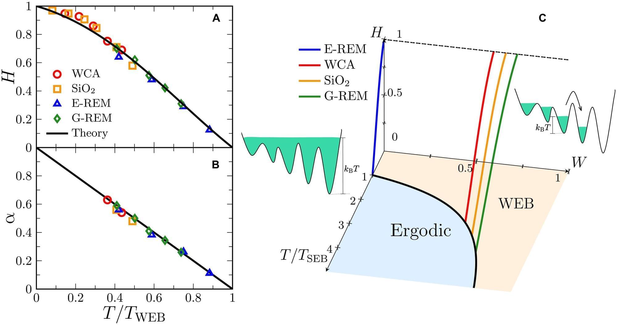
Scientists unveil universal aging mechanism in glassy materials
“Glass” has a unique and distinct meaning in physics—one that refers not just to the transparent material we associate with window glass. Instead, it refers to any system that looks solid but is not in true equilibrium and continues to change extremely slowly over time. Examples include window glass, plastics, metallic glasses, spin glasses (i.e., magnetic systems), and even some biological and computational systems.
When a liquid is cooled very quickly—a process called quenching—it doesn’t have time to organize into a crystal but becomes stuck in a disordered state far from equilibrium. Its properties—like stiffness and structure—slowly evolve through a process called “aging.”
Now, a research team from the Institute of Theoretical Physics of the Chinese Academy of Sciences has proposed a new theoretical framework for understanding the universal aging behavior of glassy materials. The study is published in the journal Science Advances.
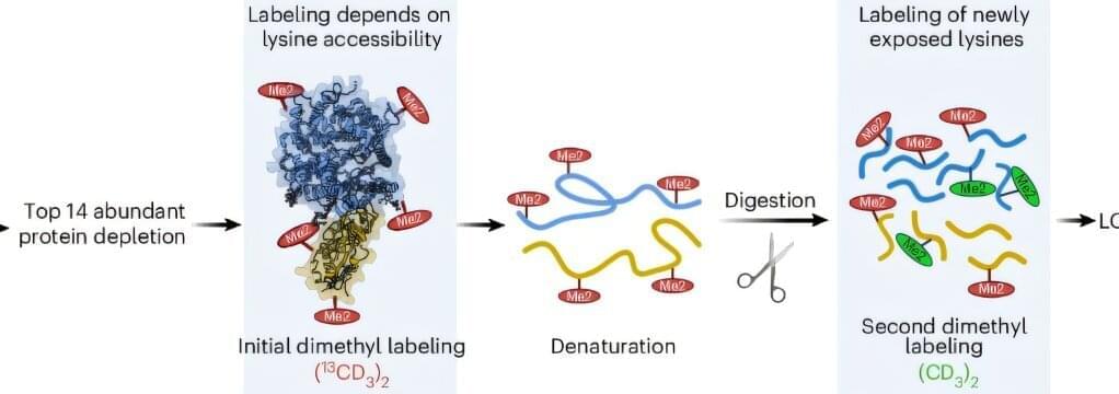
A new class of Alzheimer’s biomarkers: Why protein shape may beat protein levels
Researchers have identified a new type of blood-based biomarker test for Alzheimer’s disease that measures structural changes in proteins, providing more information on the underlying biology of the disease than standard blood tests. The findings, published in Nature Aging, also provide new insights into how Alzheimer’s disease biology may differ between males and females.
“This work introduces a fundamentally new, blood-based approach to detecting and staging Alzheimer’s disease,” said Dr. Richard Hodes, director of NIH’s National Institute on Aging (NIA). “By revealing protein structural changes associated with genetic risk, symptom severity, and sex differences—features not captured by existing biomarkers—this research could enable earlier diagnosis and more effective clinical trials.”
A biotech company just doubled the lifespan of mice without changing their diet and without editing their genes
Instead, they trained the immune system to hunt down and destroy the cells that make the body age. Then they flooded the body with fresh stem cells to rebuild what was lost.
This isn’t science fiction. It’s longevity science happening right now.
Muscle repair may hinge on a timed metabolic ‘switch,’ study suggests
Scientists at the University of California, Irvine’s School of Pharmacy & Pharmaceutical Sciences have discovered how muscle stem cells “flip a switch” to rebuild damaged muscle—a finding that could help address muscle loss linked to aging, injury and widely used weight-loss medications.
The study, published this week in Nature Metabolism, shows that muscle recovery is not just about protein or exercise. It depends on timing and how muscle cells use fuel.
Researchers learned that immediately after stress, muscle stem cells temporarily slow down energy production. Instead of burning glucose for energy, they reroute it into protective repair processes to produce antioxidants that reduce inflammation. Once repairs are complete, energy production ramps back up and new muscle fibers form and strengthen.
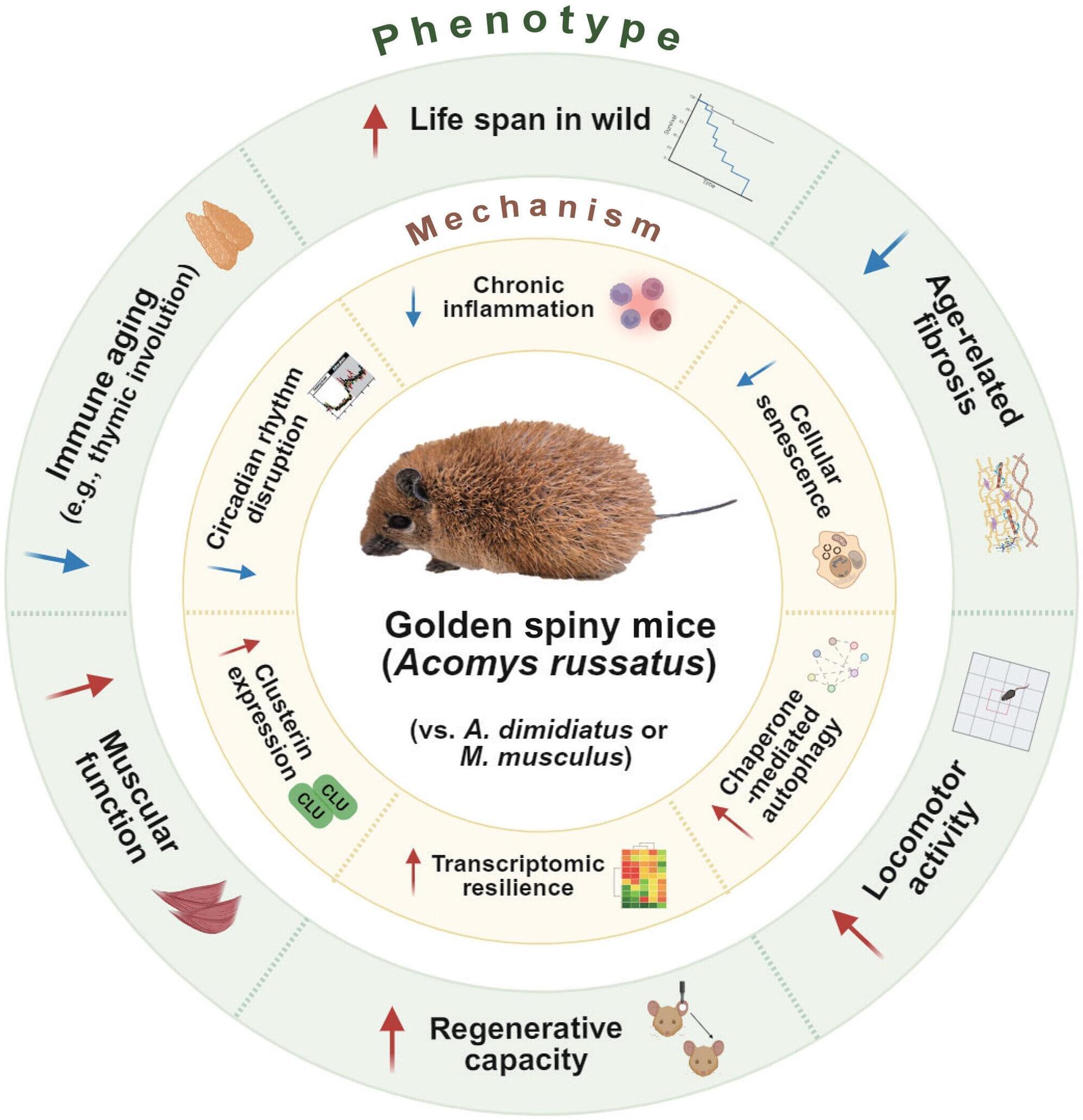
Long-living wild mouse may hold secret to healthy aging
When it comes to health, some of our animal neighbors have extraordinary advantages. Ostriches, for example, are highly resistant to viruses, while sharks rarely develop cancer. And species like naked mole rats and bowhead whales live for astonishingly long periods of time, decades and centuries, respectively.
Researchers are now starting to understand why another species—the golden spiny mouse—seems to be unhindered by the negative health effects that typically accompany aging.
Reporting in Science Advances, researchers at Yale School of Medicine (YSM) have begun to uncover how this wild mouse, native to rocky deserts in the Middle East, resists physical, cognitive, and immunological decline while living six to seven times longer than other wild mice.
Cellular Reprogramming: The Expert Roundup
Cellular reprogramming is one of the technologies most associated with longevity. The field was created in 2006, when Shinya Yamanaka showed that a cocktail of four transcription factors, commonly known as OSKM, can cause de-differentiation and massive rejuvenation of a cell, creating an iPSC (induced pluripotent stem cell). About a decade later, partial reprogramming was demonstrated in vivo, where a more subtle application of the factors led to rejuvenation without compromising the cell’s identity.
Today, this field is maturing quickly, with its first clinical trials just around the corner. Academic teams and companies are working on dozens of directions and applications. We asked four experts, all involved in reprogramming-related biotech companies, to talk about their companies’ approaches and the opportunities and bottlenecks that the field faces and to offer predictions for the near and not-so-near future.
What I find most compelling about cellular reprogramming is that it revealed aging to be, at least in part, an actively maintained biological state rather than irreversible accumulation of damage. The discovery that somatic cells retain a latent capacity to reset their epigenetic and functional identity fundamentally changed how we think about cellular plasticity, identity, and time.