
Scientists from Turkey have designed a next-generation implantable biosensor using genetically engineered E. coli for molecular-level monitoring within the body that runs on its own, wirelessly, with no external batteries required.
Category: genetics – Page 95
Taxonomy of Bacteria: Identification and Classification
We’ve been looking at bacteria for a few centuries now, so how do we categorize them? We love to classify things and put them in groups, so how does that work for bacteria? Well let’s learn about Gram-staining, antigens, other phenotypic and genotypic properties, and we will be well on our way to understanding this process!
Script by Kellie Vinal.
Watch the whole Microbiology playlist: http://bit.ly/ProfDaveMicrobio.
General Chemistry Tutorials: http://bit.ly/ProfDaveGenChem.
Organic Chemistry Tutorials: http://bit.ly/ProfDaveOrgChem.
Biochemistry Tutorials: http://bit.ly/ProfDaveBiochem.
Biology/Genetics Tutorials: http://bit.ly/ProfDaveBio.
Anatomy & Physiology Tutorials: http://bit.ly/ProfDaveAnatPhys.
Biopsychology Tutorials: http://bit.ly/ProfDaveBiopsych.
Immunology Tutorials: http://bit.ly/ProfDaveImmuno.
History of Drugs Videos: http://bit.ly/ProfDaveHistoryDrugs.
EMAIL► [email protected].
PATREON► / professordaveexplains.
Check out \.
Who’s Really Winning At Longevity? (Featuring @Unaging.Crissman.Loomis)
Join us on Patreon! https://www.patreon.com/MichaelLustgartenPhD
Discount Links/Affiliates:
Blood testing (where I get the majority of my labs): https://www.ultalabtests.com/partners/michaellustgarten.
At-Home Metabolomics: https://www.iollo.com?ref=michael-lustgarten.
Use Code: CONQUERAGING At Checkout.
Clearly Filtered Water Filter: https://get.aspr.app/SHoPY
Epigenetic, Telomere Testing: https://trudiagnostic.com/?irclickid=U-s3Ii2r7xyIU-LSYLyQdQ6…M0&irgwc=1
Use Code: CONQUERAGING
NAD+ Quantification: https://www.jinfiniti.com/intracellular-nad-test/
Continue reading “Who’s Really Winning At Longevity? (Featuring @Unaging.Crissman.Loomis)” | >
Scientists shed new light on the shared genetic basis of psychiatric disorders
New research uncovers how shared genes contribute to various psychiatric disorders. This breakthrough highlights the importance of pleiotropic genes—those affecting multiple conditions—and offers new avenues for mental health treatment development.

Children with better musical skills may benefit from a prolonged window of brain plasticity
Children who excel at keeping the beat may possess brains that mature more slowly, extending their capacity for learning. A new longitudinal twin study indicates that this prolonged development is shaped by both genetic predispositions and musical engagement.
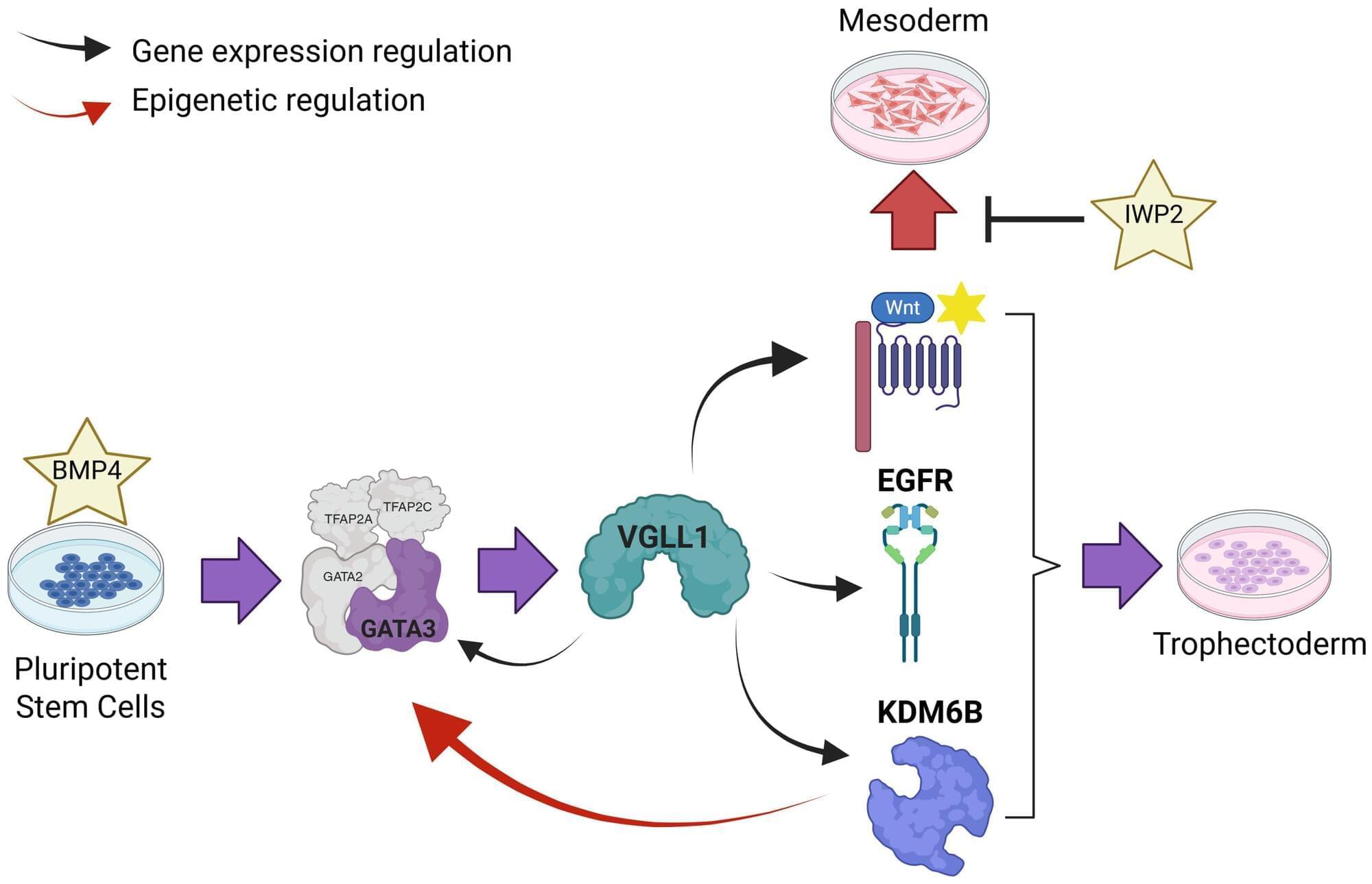
Researchers uncover the earliest stages of human placenta formation
A gene that turns on very early in embryonic development could be key to the formation of the placenta, which provides the developing fetus with what it needs to thrive during gestation.
The placenta provides all of the nutrition, oxygen and antibodies that a developing human fetus needs to thrive throughout gestation. The temporary organ begins to form within six to 12 days after conception, just as the embryo implants itself in the lining of the uterus. Failure of the placenta to form correctly is the second leading cause of miscarriage during early pregnancy, after genetic abnormalities of the fetus that are incompatible with life.
However, the initial stages of placental formation have remained a mystery due to ethical considerations and technical constraints on studying the process in humans.
Machine learning reveals how disordered protein regions contribute to cancer-causing condensates
Fusion oncoproteins arise when a gene fuses with another gene and acquires new abilities. Such abilities can include the formation of biomolecular condensates, “droplets” of concentrated proteins, DNA or RNA.
The abnormal molecular condensates formed by fusion oncoproteins can disrupt cellular functions and drive cancer development, but the specific protein features behind this process remain unclear.
Scientists at St. Jude Children’s Research Hospital studied intrinsically disordered regions, unstructured protein segments that are often involved in condensate formation, to determine if they drive fusion oncoproteins to form condensates. They trained a machine learning model, called IDR-Puncta ML, with experimental data from intrinsically disordered regions in fusion oncoproteins to predict the behavior of other such regions.

One Dose of This Gene Editor Could Defeat a Host of Genetic Diseases Suffered by Millions
The gene-editing tool takes on “nonsense mutations” that are involved in roughly 30 million genetic diseases.
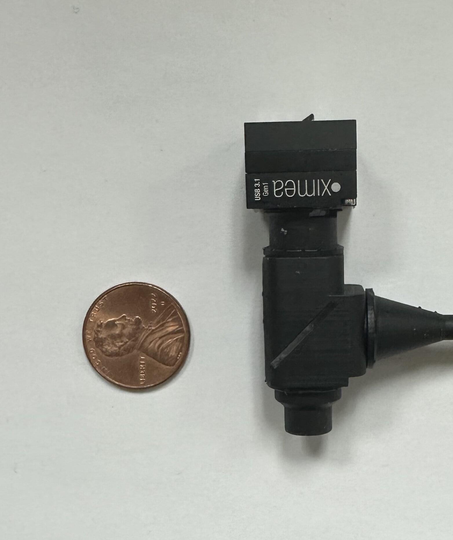
Miniature microscope captures real-time voltage signals in awake animals
Researchers have built a tiny, lightweight microscope that captures neuron activity with unprecedented speed that can be used in freely moving animals. The new tool could give scientists a more complete view of how brain cells process information during natural behavior.
The microscope is designed to image genetically encoded voltage indicators —fluorescent dyes that rapidly change brightness when a neuron fires—through a small window in the skull while the animal is awake.
“Unlike most miniature microscopes that track slower calcium signals, ours captures electrical spikes at hundreds of frames per second,” said Emily Gibson from the University of Colorado Anschutz Medical Campus. “This makes it possible to capture the moment a neuron fires as well as the quieter signals that build up inside neurons before firing.”
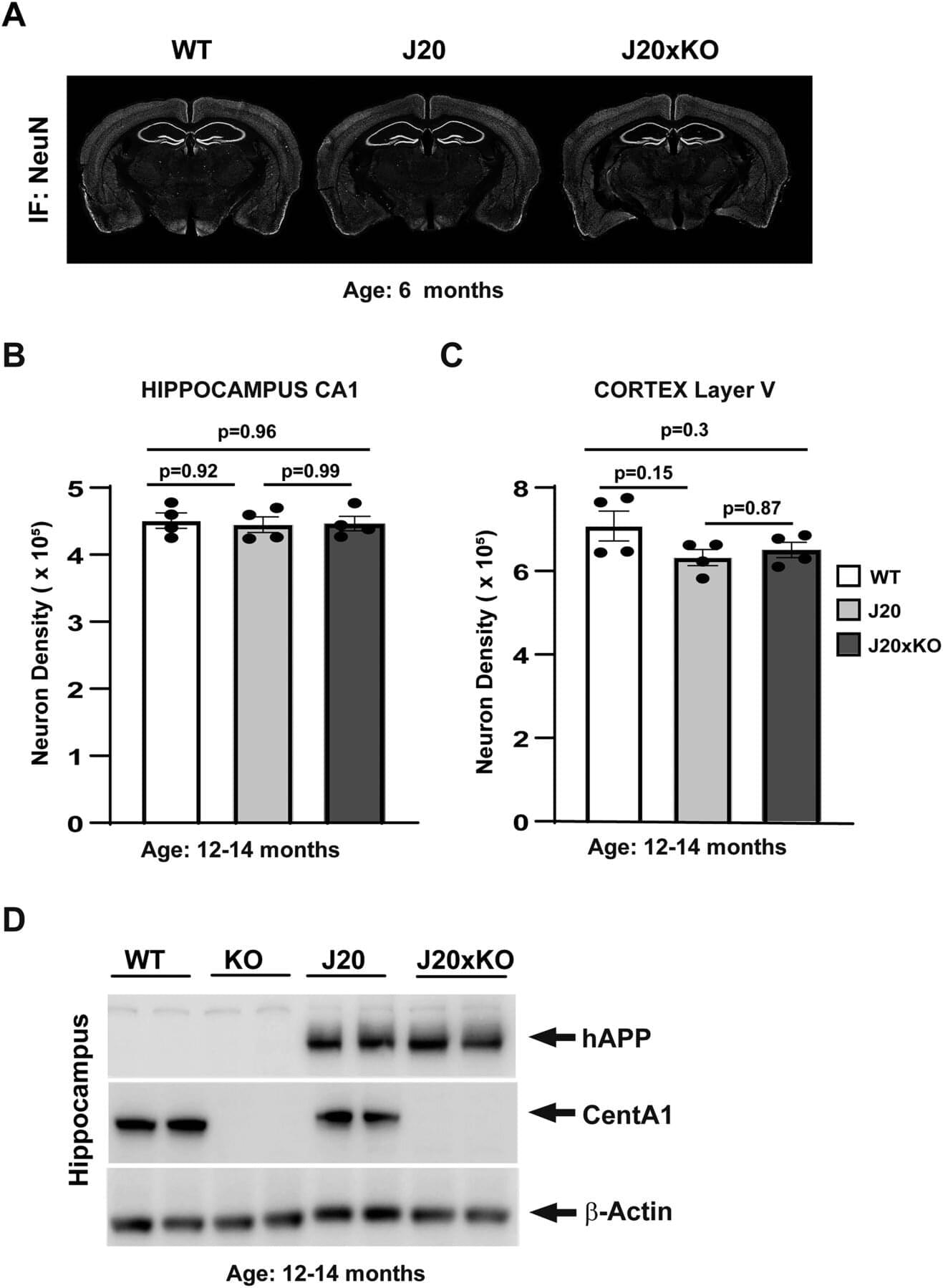
Loss of brain protein eases Alzheimer’s symptoms and brain damage in mice
New research published in the journal eNeuro examined whether eliminating a protein that is elevated in the brains of those with Alzheimer’s could prevent or reduce damage and behavioral symptoms in a mouse model of Alzheimer’s disease.
“Previous work from our research team and others found evidence that a specific protein named Centaurin-α1 is involved in the progression of Alzheimer’s damage within neurons,” explained lead author of the study, Dr. Erzsebet Szatmari. “To confirm the role of this protein and see if it might be a good therapeutic target, we tested whether genetically removing it would prevent or slow disease progression in a mouse model of the disease.”
The scientists used a well-characterized model of Alzheimer’s disease in mice. The disease model (called J20) contains two genetic mutations associated with rare familial variants of Alzheimer’s disease. These animals develop changes in brain tissue and behavioral deficits characteristic of many symptoms seen in human Alzheimer’s disease, including neuroinflammation, accumulation of neuronal plaques, synapse loss, and impairments in spatial memory and learning.