Alzheimer’s disease starts with a sticky protein called amyloid beta that builds up into plaques in the brain, setting off a chain of events that results in brain atrophy and cognitive decline. Microglia, immune cells that reside in the brain, are responsible for removing brain waste but can become dysfunctional when overwhelmed in the context of neurodegenerative disease.
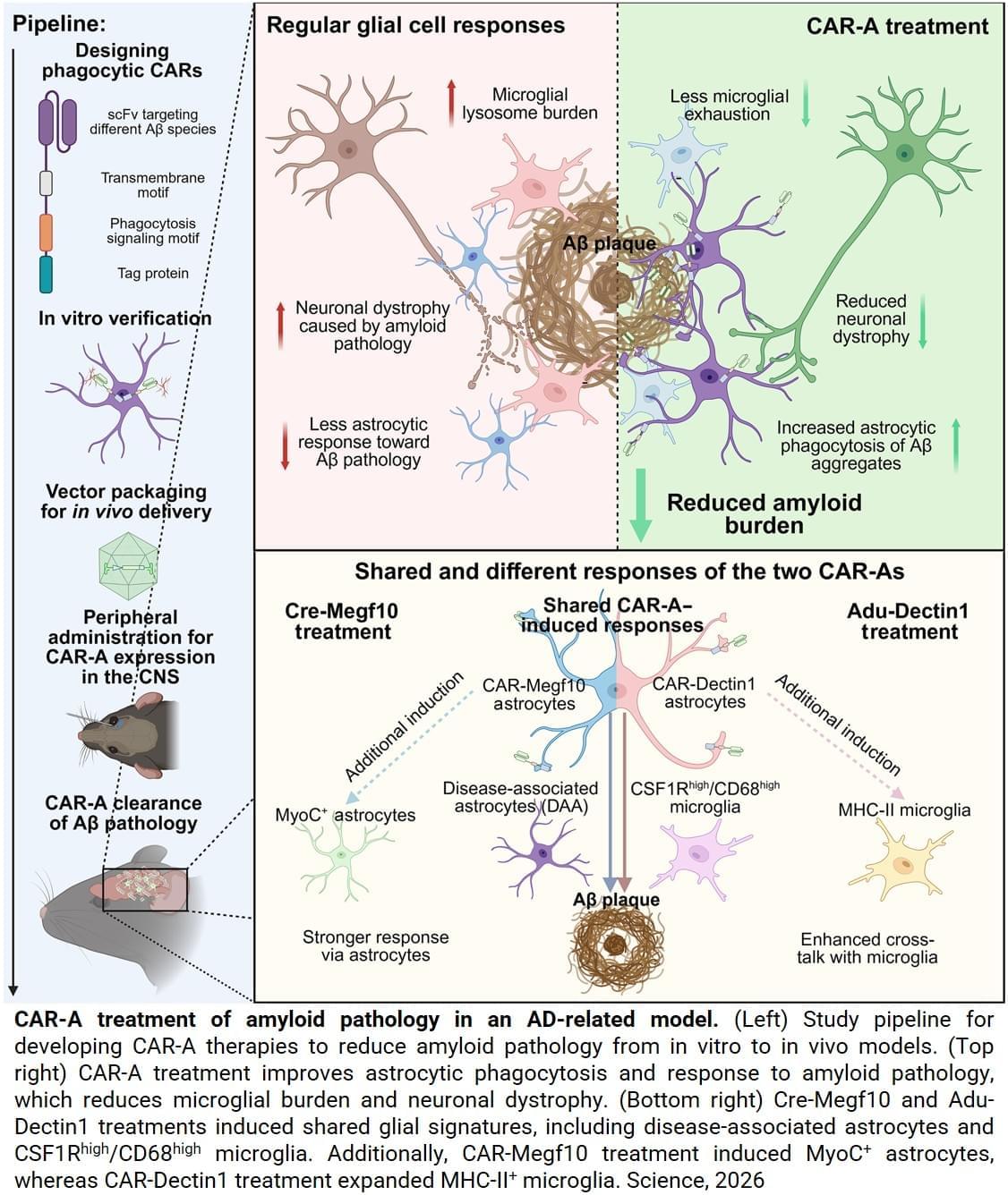
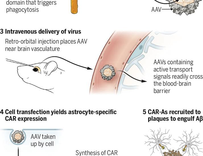
To reduce the cleaning burden on microglia, first author transformed astrocytes, the most abundant cell type in the brain, into amyloid-cleaning machines. The author custom-designed and delivered a gene to astrocytes that codes for the chimeric antigen receptor (CAR) via a harmless virus injected into mice. The CAR, now present on the surface of astrocytes, enabled the cells to capture and engulf amyloid beta proteins. With their newly acquired ability, the astrocytes — generally responsible for keeping the brain tidy — concentrated their efforts on only cleaning amyloid beta plaques in mice prone to its buildup.
Mice carrying genetic mutations that increase people’s risk of developing Alzheimer’s disease develop amyloid beta plaques that saturate the brain by six months of age. The author injected two groups of mice with the virus carrying the CAR-expressing gene: young mice before they developed plaques and older mice with brains saturated with plaques, then, waited three months.
As the younger mice aged, the CAR-astrocytes prevented amyloid beta plaque development. At nearly six months of age, when untreated mice normally have brains saturated with harmful plaques, brains of treated mice were plaque-free. Meanwhile, older mice with plaque-saturated brains at the time of treatment saw a 50% reduction in the amount of amyloid beta plaques compared to mice receiving an injection of a virus lacking the CAR gene.
The researchers have filed a patent related to the approach used to engineer CAR-astrocytes.
“Consistent with the antibody drug treatments, this new CAR-astrocyte immunotherapy is more effective when given in the earlier stages of the disease,” said a co-author on the paper. “But where it differs, and where it could make a difference in clinical care, is in the single injection that successfully reduced the amount of harmful brain proteins in mice.” ScienceMission sciencenewshighlights.