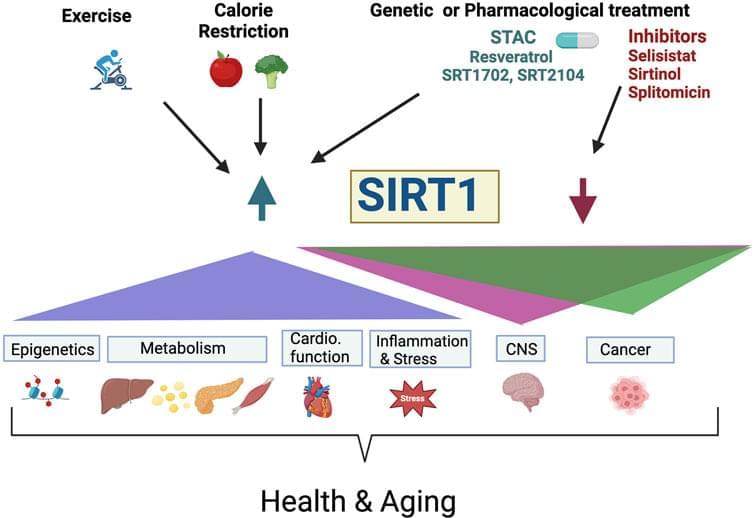
Toggle light / dark theme

In a new study published in Nature, researchers have developed a breakthrough technique called spatial transcriptomics, which allows scientists to map tumors non-invasively and at an unprecedented resolution depth. For the first time, researchers have created a three-dimensional map of a whole prostate to an unprecedented resolution, including areas of healthy and cancerous cells. Surprisingly, the study revealed that individual prostate tumors contain a range of genetic variations, which until this point were unknown.

“We have never had this level of resolution available before, and this new approach revealed some surprising results,” said Alastair Lamb of Oxford’s Nuffield Department of Surgical Sciences, who jointly led the study.

In recent years, ribonucleic acid (RNA) has emerged as a powerful tool for the development of novel therapies. RNA is used to copy genetic information contained in our hereditary material, the deoxyribonucleic acid (DNA), and then serves as a template for building proteins, the building blocks of life. Delivery of RNA into cells remains a major challenge for the development of novel therapies across a broad range of diseases. Researchers at the Max Planck Institute of Molecular Cell Biology and Genetics (MPI-CBG) in Dresden together with researchers from the global biopharmaceutical company AstraZeneca have investigated where and how mRNA is delivered inside the cell. They found that mRNA uses an unexpected entry door. Their results provide novel insights into the development of RNA therapeutics towards efficient delivery and lower dosages.

DNA () contains the required for the development and maintenance of life. This information is communicated by messenger (mRNA) to make proteins. mRNA-based therapeutics have the potential to address unmet needs for a wide variety of diseases, including cancer and cardiovascular disease. mRNA can be delivered to cells to trigger the production, degradation or modification of a target protein, something impossible with other approaches. A key challenge with this modality is being able to deliver the mRNA inside the cell so that it can be translated to make a protein. mRNA can be packed into lipid nanoparticles (LNPs)—small bubbles of fat—that protect the mRNA and shuttle it into cells. However, this process is not simple, because the mRNA has to pass the membrane before it can reach its site of action in the cell interior, the cytoplasm.

Researchers in the team of MPI-CBG director Marino Zerial are experts in visualizing the cellular entry routes of molecules in the cell, such as mRNA with high-resolution microscopes. They teamed up with scientists from AstraZeneca who provided the researchers with lipid nanoparticle prototypes that they had developed for therapeutic approaches to follow the mRNA inside the cell. The study is published in the Journal of Cell Biology.

Nanoengineers at the University of California San Diego have developed a new and potentially more effective way to deliver messenger RNA (mRNA) into cells. Their approach involves packing mRNA inside nanoparticles that mimic the flu virus—a naturally efficient vehicle for delivering genetic material such as RNA inside cells.

The new mRNA nanoparticles are described in a paper published recently in the journal Angewandte Chemie International Edition.

The work addresses a major challenge in the field of drug delivery: Getting large biological drug molecules safely into and protecting them from organelles called endosomes. These tiny acid-filled bubbles inside the cell serve as barriers that trap and digest large molecules that try to enter. In order for biological therapeutics to do their job once they are inside the cell, they need a way to escape the endosomes.

Human Augmentation Examples. What are examples of augmentations to the human body? Should we allow such augmentation methods and technologies? What are dangers, and risks involved? How can society benefit from these developments?

On Brave New World conference 2020 I gave this webinar with the title: ‘The Human Body. The Next Frontier’.

Please leave a comment if you like the video or when you have a question!

Content:

Specific proteins in prokaryotes detect viruses in unexpectedly direct ways.

Bacteria use a variety of defense strategies to fight off viral infection. STAND ATPases in humans are known to respond to bacterial infections by inducing programmed cell death in infected cells. Scientists predict that many more antiviral weapons will be discovered in the microbial world in the future. Scientists have discovered a new unexplored microbial defense system in bacteria.

Researchers uncovered specific proteins in prokaryotes (bacteria and archaea) that detect viruses in unexpectedly direct ways, recognizing critical parts of the viruses and causing the single-celled organisms to commit suicide to stop the infection within a microbial community, according to a press release published in the official website of the Massachusetts Institute of Technology (MIT) on Thursday.

The discovery was made by a team of scientists led by researchers at the Broad Institute of MIT and Harvard and the McGovern Institute for Brain Research at MIT.

“This work demonstrates a remarkable unity in how pattern recognition occurs across very different organisms,” said Feng Zhang, senior author and James, and Patricia Poitras Professor of Neuroscience at MIT.

Circa 2015


“Their work has provided fundamental knowledge of how a living cell functions and is, for instance, used for the development of new cancer treatments,” the Royal Swedish Academy of Sciences said.

Thousands of alterations to a cell’s genome occur every day due to spontaneous changes and damage by radiation, free radicals and carcinogenic substances — yet DNA remains astonishingly intact.

To keep genetic materials from disintegrating, a range of molecular systems monitor and repair DNA, in processes that the three award-winning scientists helped map out.

This extensive research actually details the possibility of unknown origins of these elongated human skulls which many think to this day are some form of exterrestial in origin or at the very least unknown in origin which actually nearly uproots most know origin stories.


Modern European genetic structure demonstrates strong correlations with geography, while genetic analysis of prehistoric humans has indicated at least two major waves of immigration from outside the continent during periods of cultural change. However, population-level genome data that could shed light on the demographic processes occurring during the intervening periods have been absent. Therefore, we generated genomic data from 41 individuals dating mostly to the late 5th/early 6th century AD from present-day Bavaria in southern Germany, including 11 whole genomes (mean depth 5.56×). In addition we developed a capture array to sequence neutral regions spanning a total of 5 Mb and 486 functional polymorphic sites to high depth (mean 72×) in all individuals. Our data indicate that while men generally had ancestry that closely resembles modern northern and central Europeans, women exhibit a very high genetic heterogeneity; this includes signals of genetic ancestry ranging from western Europe to East Asia. Particularly striking are women with artificial skull deformations; the analysis of their collective genetic ancestry suggests an origin in southeastern Europe. In addition, functional variants indicate that they also differed in visible characteristics. This example of female-biased migration indicates that complex demographic processes during the Early Medieval period may have contributed in an unexpected way to shape the modern European genetic landscape. Examination of the panel of functional loci also revealed that many alleles associated with recent positive selection were already at modern-like frequencies in European populations ∼1,500 years ago.

Realistic and complex models of brain cells, developed at Cedars-Sinai with support from our scientists and our #openscience data, could help answer questions a… See more.


Cedars-Sinai investigators have created bio-realistic and complex computer models of individual brain cells—in unparalleled quantity.

Their research, published today in the peer-reviewed journal Cell Reports, details how these models could one day answer questions about neurological disorders—and even human intellect—that aren’t possible to explore through biological experiments.

“These models capture the shape, timing and speed of the electrical signals that neurons fire in order to communicate with each other, which is considered the basis of brain function,” said Costas Anastassiou, PhD, a research scientist in the Department of Neurosurgery at Cedars-Sinai, and senior author of the study. “This lets us replicate brain activity at the single-cell level.”

A team at the University of California, Irvine, has identified a signaling molecule that potently stimulates hair growth.

A signaling molecule known as SCUBE3, which was discovered by researchers at the University of California, Irvine, has the potential to cure androgenetic alopecia, a prevalent type of hair loss in both women and men.

The research, which was recently published in the journal Developmental Cell, uncovered the precise mechanism by which the dermal papilla cells, specialized signal-producing fibroblasts found at the bottom of each hair follicle, encourage new development. Although the critical role dermal papilla cells play in regulating hair growth is widely established, the genetic basis of the activating chemicals involved is little understood.