A new study has unveiled when chronic myeloid leukaemia, a type of cancer that affects the blood and bone marrow, arises in life and how fast it grows. Researchers reveal explosive growth rates of cancerous cells years before diagnosis and variation in these rates of growth between patients. Such rapid growth rates had previously not been observed in most other cancers.
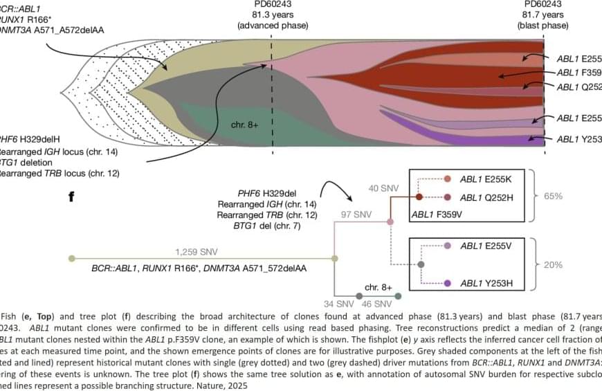
Researchers used whole genome sequencing to study when BCR::ABL1 – an abnormal fusion of the different genes called BCR and ABL1, which is known to cause chronic myeloid leukaemia. The team investigated when BCR::ABL1 first arises in a blood cell and how quickly these cells with this genetic change then multiply and expand to lead to a diagnosis of a type of leukaemia.
The research, published in Nature, contributes to the scientific understanding of how strong this abnormal fusion gene is in its ability to drive cancer.


