Toggle light / dark theme

Join us on Patreon! https://www.patreon.com/MichaelLustgartenPhD

Discount Links:
At-Home Metabolomics: https://www.iollo.com?ref=michael-lustgarten.
Use Code: CONQUERAGING At Checkout.

NAD+ Quantification: https://www.jinfiniti.com/intracellular-nad-test/
Use Code: ConquerAging At Checkout.

Epigenetic Testing: https://trudiagnostic.com/?irclickid=U-s3Ii2r7xyIU-LSYLyQdQ6…M0&irgwc=1

Insects, with their remarkable ability to undergo complete metamorphosis, have long fascinated scientists seeking to understand the underlying genetic mechanisms governing this transformative process.

Now, a recent study conducted by the Institute for Evolutionary Biology (IBE, CSIC-UPF) and the IRB Barcelona has shed light on the crucial role of three genes – Chinmo, Br-C and E93 – in orchestrating the stages of insect development. Published in eLife, this research provides valuable insights into the evolutionary origins of metamorphosis and sheds new light on the role of these genes in growth, development and cancer regulation [1].

Longevity. Technology: Chinmo might sound like a Pokémon character, but the truth is much more interesting. Conserved throughout the evolution of insects, scientists think it, and the more conventionally-named Br-C and E93, could play a key role in the evolution of metamorphosis, acting as the hands of the biological clock in insects. A maggot is radically different from the fly into which it changes – could understanding and leveraging the biology involved one day allow us to change cultured skin cells into replacement organs or to stop tumors in their early stages of formation? No, Dr Seth Brundle, you can buzz off.

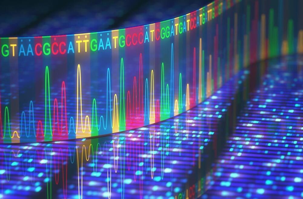
Aleksandra Radenovic, head of the Laboratory of Nanoscale Biology in the School of Engineering, has worked for years to improve nanopore technology, which involves passing a molecule like DNA through a tiny pore in a membrane to measure an ionic current. Scientists can determine DNA’s sequence of nucleotides—which encodes genetic information—by analyzing how each one perturbs this current as it passes through. The research has been published in Nature Nanotechnology.

Currently, the passage of molecules through a and the timing of their analysis are influenced by random physical forces, and the rapid movement of molecules makes achieving high analytical accuracy challenging. Radenovic has previously addressed these issues with optical tweezers and viscous liquids. Now, a collaboration with Georg Fantner and his team in the Laboratory for Bio-and Nano-Instrumentation at EPFL has yielded the advancement she’s been looking for—with results that could go far beyond DNA.

“We have combined the sensitivity of nanopores with the precision of scanning ion conductance microscopy (SICM), allowing us to lock onto specific molecules and locations and control how fast they move. This exquisite control could help fill a big gap in the field,” Radenovic says. The researchers achieved this control using a repurposed state-of-the-art scanning ion conductance microscope, recently developed at the Lab for Bio-and Nano-Instrumentation.

Join us on Patreon! https://www.patreon.com/MichaelLustgartenPhD

Discount Links:
Telomere Length, Epigenetic Testing: https://trudiagnostic.com/?irclickid=U-s3Ii2r7xyIU-LSYLyQdQ6…M0&irgwc=1
Use Code: CONQUERAGING

NAD+ Quantification: https://www.jinfiniti.com/intracellular-nad-test/
Use Code: ConquerAging At Checkout.

At-Home Metabolomics: https://www.iollo.com?ref=michael-lustgarten.

A human cell harbors roughly 2 meters of DNA, encompassing the essential genetic information of an individual. If one were to unwind and stretch out all the DNA contained within a single person, it would span a staggering distance—enough to reach the sun and back 60 times over. In order to manage such an astounding volume of biological information, the cell compacts its DNA into tightly packed chromosomes.

“Imagine DNA as a piece of paper upon which all our is written,” says Minke A.D. Nijenhuis, co-corresponding author. “The paper is folded into a very tight structure in order to fit all of that information into a small cell nucleus. To read the information, however, parts of the paper have to be unfolded and then refolded. This spatial organization of our genetic code is a central mechanism of life. We therefore wanted to create a methodology that allows researchers to engineer and study the compaction of double-stranded DNA.”

Natural DNA is often double-stranded: one strand to encode the genes and one backup strand, intertwined in a . The double helix is stabilized by Watson-Crick interactions, which allow the two strands to recognize and pair with one another. Yet there exists another, lesser-known class of interactions between DNA. These so-called normal or reverse Hoogsteen interactions allow a third strand to join in, forming a beautiful triple helix (Figure 1).

Synthetic human embryos – derived from stem cells without the need for eggs or sperm – have been created for the first time, scientists say. The structures represent the very earliest stages of human development, which could allow for vital studies into disorders like recurrent miscarriage and genetic diseases. But questions have been posed about the legal and ethical implications, as the pace of scientific discovery outstrips the legislation.

The breakthrough was reported by the Guardian newspaper following an announcement by Professor Magdalena Żernicka-Goetz, a developmental biologist at the University of Cambridge and Caltech, at the 2023 annual meeting of the International Society for Stem Cell Research. The findings have not yet been published in a peer-reviewed paper.

It’s understood that the synthetic structures model the very beginnings of human development. They do not yet contain a brain or heart, for example, but comprise the cells that would be needed to form a placenta, yolk sac, and embryo. Żernicka-Goetz told the conference that the structures have been grown to just beyond the equivalent of 14 days of natural gestation for a human embryo in the womb. It’s not clear whether it would be possible to allow them to mature any further.

Healthy cells work hard to maintain the integrity of our DNA, but occasionally, a chromosome can get separated from the others and break apart during cell division. The tiny fragments of DNA then get reassembled in random order in the new cell, sometimes producing cancerous gene mutations.

This chromosomal shattering and rearranging is called “chromothripsis” and occurs in the majority of human cancers, especially cancers of the bones, brain and fatty tissue. Chromothripsis was first described just over a decade ago, but scientists did not understand how the floating pieces of DNA were able to be put back together.

In a study published in Nature, researchers at University of California San Diego have answered this question, discovering that the shattered DNA fragments are actually tethered together. This allows them to travel as one during and be re-encapsulated by one of the new daughter cells, where they are reassembled in a different order.

Researchers at the University of Houston are working to make T-cell immunotherapy safer, developing a tool called CrossDome, which uses a combination of genetic and biochemical information to predict if T-cell immunotherapies might mistakenly attack healthy cells.

T-cell based immunotherapies hold tremendous potential in the fight against cancer and , thanks to their capacity to specifically target diseased cells, including cancer metastasis. Nevertheless, this potential has been tempered with safety concerns regarding the possible recognition of unknown off targets displayed by .

In one case, scientists created special T-cells that were supposed to target a protein found in a type of skin cancer called melanoma. However, these T-cells also ended up attacking a different protein found in the heart cells of some patients. This caused severe damage to the heart.