Be kind to yourself this year. Using Zocdoc is FREE — visit my sponsor https://zocdoc.com/quinnsideas to find and instantly book an appointment with a top-rated, in-network doctor today.
Imagine a civilization reaches something like a Type II level, advanced enough to move through interstellar space and keep large populations alive for generations. At that stage, the challenge is developing ships that can cross the void, and also making sure the people inside them can survive radiation, isolation, and extreme travel times. That could mean heavy genetic engineering before the journey begins, changing bone density, metabolism, resistance to disease, tolerance for low gravity, or even sensory systems and respiration. But when they finally arrive, they may still find that the planet is wrong for them, maybe the air is toxic, the gravity is crushing, the temperatures are extreme, or the native chemistry is incompatible with human biology.
At that point, they face two paths. One is terraforming, which means trying to remake an entire planet into something closer to Earth. That could involve thickening or thinning an atmosphere, warming a frozen world, cooling a hot one, importing water, altering soil chemistry, introducing engineered microbes, building orbital mirrors or shades, and managing the planet for centuries or even millennia. The scale of that project is absurdly expensive, not just in money but in energy, infrastructure, labor, time, and raw materials. You are not changing a city or even a continent, you are trying to rewrite a whole world.
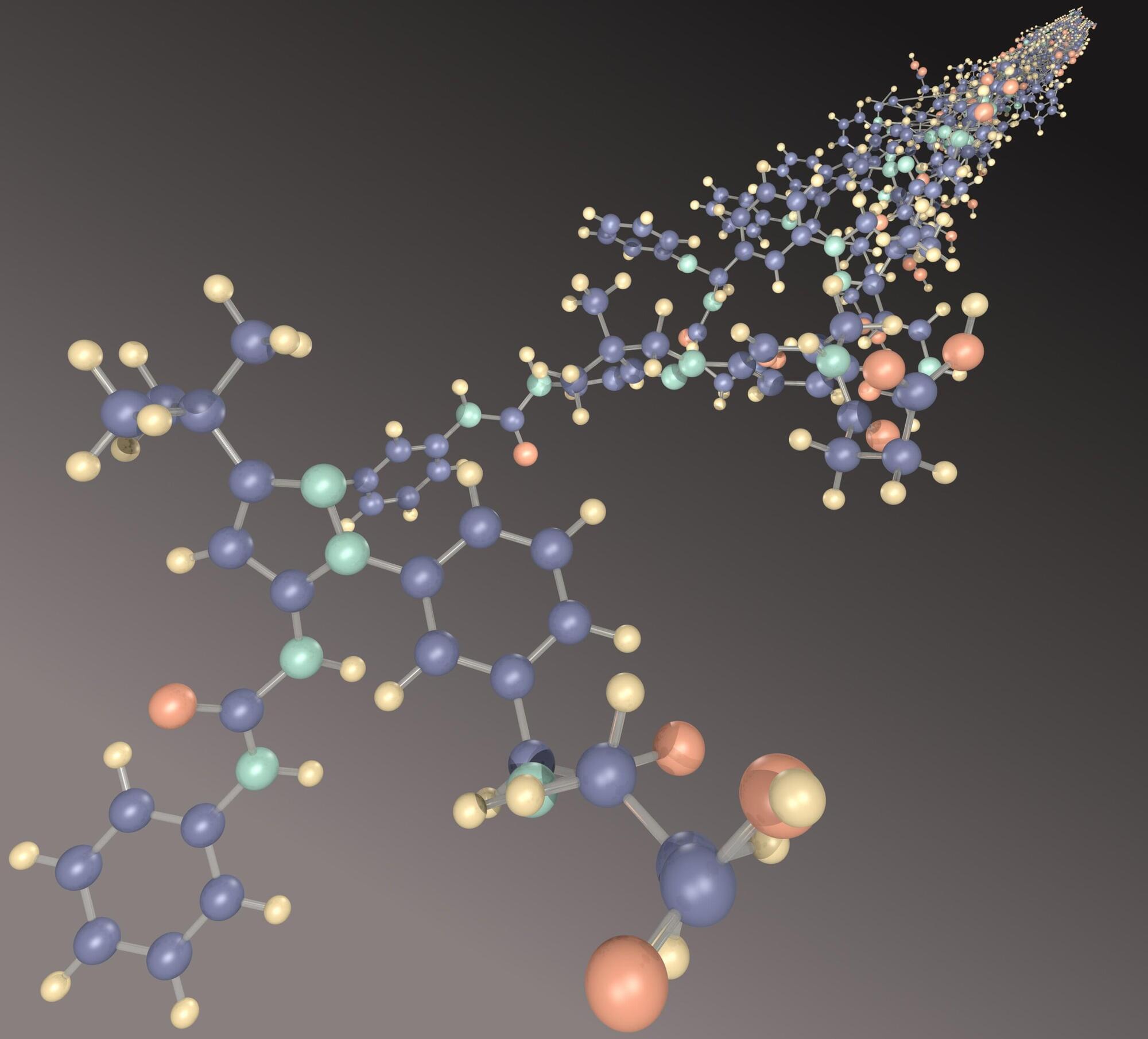
The other option is pantropy. Instead of forcing the planet to become Earth-like, the colonists change themselves to fit the planet. They might alter their lungs to breathe a different atmospheric mix, redesign their skin to handle harsher radiation, reduce their size for lower resource use, strengthen their bodies for higher gravity, or even become something so biologically different that they no longer look fully human. That is the core idea of pantropy, adapting the colonists to the world rather than adapting the world to the colonists.
The term was coined by James Blish, and he used it in connection with the stories collected in The Seedling Stars, especially “Surface Tension.” which was first published in 1952 in Galaxy Science Fiction.
Get 50% off Nebula (get access, early videos, exclusives): https://go.nebula.tv/quinnsideas.