A plastic surgeon and burn specialist, Fiona Wood, developed a spray-on skin method to treat burns without scarring.


The experimental drug combo dasatinib and quercetin (known for short as D+Q) is one of the most promising anti-aging therapies being developed right now.
It is not yet approved for human use, but some scientists think it has the potential to fight disease by improving how our systems clear out worn-down cells.
According to a new study, however, there might be a big problem with D+Q.
Telomeres are regarded as key markers of cellular ageing and physiological state. Oxidative stress, which can accelerate telomere shortening, is thought to increase during energetically demanding processes such as bird migration. However, their study in the context of migratory behaviour is limited. Here we compared telomere length (TL) and mitochondrial DNA copy number (mitoDNAcn) between migratory and resident Eurasian blackbirds on the island of Helgoland, a migratory stopover site. Contrary to expectations, we found migrants had longer TLs despite similar mitochondrial densities. These findings reinforce the idea that migratory individuals may possess specific physiological adaptations, such as enhanced antioxidant defences, that help preserve telomere integrity.

Giving AI a classic psychological test reveals an inherent weakness in LLM decision-making abilities. Suketu Patel and colleagues explored how transformer-based machine attention differs from human attention by testing AI models on the “Stroop task,” in which words for colors are printed in colored ink, and participants are asked to name the ink color of each word while ignoring its meaning.
The findings are published in the journal PNAS Nexus.
The task is clinically used to assess executive control, especially a person’s ability to inhibit an automatic response. Although humans generally take longer to answer correctly when words and colors are mismatched than when they match, they can still perform stably and with high accuracy even on long word lists.
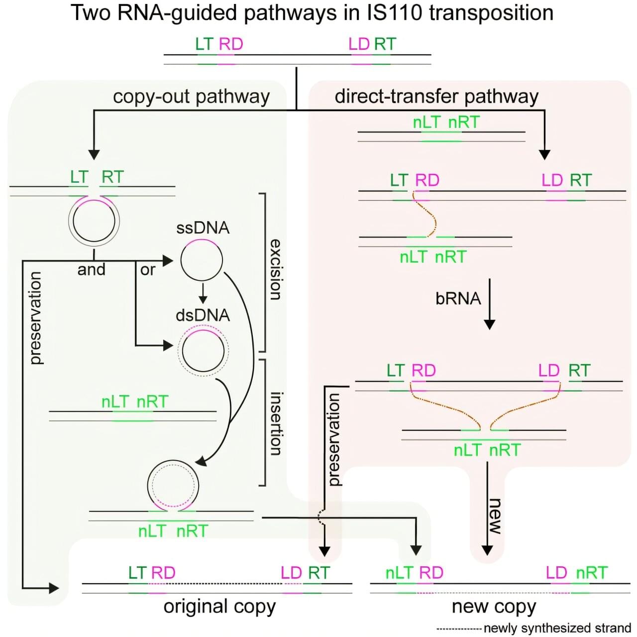
IS110 transposons are a large, diverse family of bacterial insertion sequences (IS elements)—small, mobile DNA elements that can move from one genomic location to another. They have recently attracted broad interest due to the finding that some of these transposons use a bridge RNA (bRNA) to recognize both donor DNA and target DNA.
Upon this discovery, researchers hoped that bRNA-guided transposon systems could offer a genome-editing strategy distinct from classical CRISPR-Cas nucleases and thereby enable programmable DNA integration. However, it remained unclear how IS110 elements insert donor DNA into target sites and whether these elements rely on one or multiple reaction pathways.
Now, a new study led by Xue Chaoyou from the Tianjin Institute of Industrial Biotechnology of the Chinese Academy of Sciences, in collaboration with Lou Huiqiang at China Agricultural University and RAO Shuquan from the Chinese Academy of Medical Sciences, answers these questions by showing that RNA-guided IS110 transposons use two mechanistically distinct pathways to mobilize DNA.
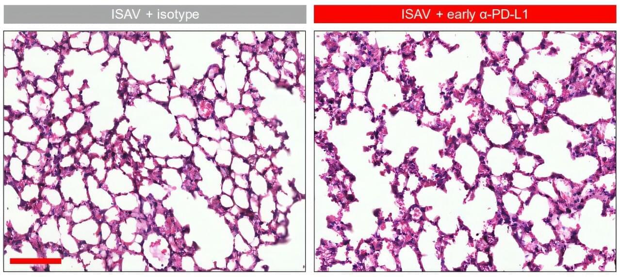
A new study led by researchers at The University of Texas MD Anderson Cancer Center has shown that early administration of immunotherapy with standard antifungal treatment improved outcomes and largely alleviated immune system paralysis caused by fungal lung infections in preclinical models. These findings could herald new clinically relevant strategies for treating a variety of life-threatening invasive fungal pneumonias, which disproportionately affect immunocompromised cancer patients.
The study, published in the Proceedings of the National Academy of Sciences, was led by Sebastian Wurster, M.D., assistant professor, and Dimitrios P. Kontoyiannis, M.D., Ph.D., professor, both of Infectious Diseases, Infection Control and Employee Health.
“Despite an expanded arsenal of antifungal treatments, immune system dysfunction is still a major cause of failure when treating infections, with significantly high morbidity and mortality rates associated with pneumonias caused by opportunistic molds. There is an urgent need for adjunct immune-enhancing therapies to improve outcomes,” Kontoyiannis said. “Our research shows that adding an immune checkpoint inhibitor to antifungal treatments is helpful in experimental mold pneumonias, especially when given early.”


Transient forebrain ischemia is associated with selective neuronal vulnerability and persistent memory deficit. This study compares functional outcome and morphological changes in rats subjected to post-ischemic CA1 or hilus/dentate gyrus region hippocampal fetal transplantation. Ischemia was produced by bilateral common carotid artery occlusion with hypotension. Fetal hippocampal neurons were transplanted into both sides of the CA1 or hilus/dentate gyrus region of the dorsal hippocampus, 1 week post-ischemia. Four weeks post transplantation, the rats underwent behavioral testing for 5 consecutive days using the water maze trial. All animals were perfusion fixed for morphological studies. Transplants in the CA1 region of the dorsal hippocampus were associated with memory and morphological recovery, while grafts placed into the hilus/dentate gyrus region of the dorsal hippocampus were not. Similarly, neurons transplanted in the CA1 region of the dorsal hippocampus were morphologically similar to CA1 pyramidal cell neurons and stained positive with calbindin D(28k). In contrast the grafts transplanted into the hilus/dentate gyrus region of the dorsal hippocampus were morphologically heterogeneous and staining with calbindin D(28k) was not as robust. Post-ischemic transplantation in the CA1 region of the dorsal hippocampus is effective in improving memory and morphological function.