Modern fluorescence microscopy can generate images of living cells as stunning to look at as they are informative to study. For techniques like fluorescence lifetime imaging microscopy (FLIM), those images provide a window into cell metabolism, helping scientists study cancer treatment, autoimmune disease and more.
But for these researchers, the image is just the beginning. To draw any biological insights, researchers need to guide massive amounts of data through a maze of software analysis tools and scripts, ensuring careful quality checks throughout the journey.
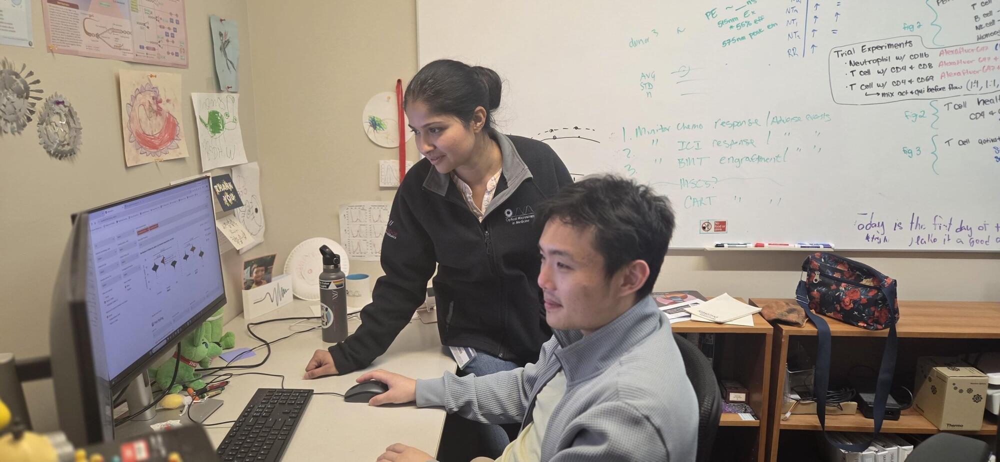
Morgridge Institute for Research scientists in the Melissa Skala Lab are tackling this challenge head-on. They have developed a new open-source, user-friendly data analysis platform, FLIM Playground, designed to make FLIM analysis easier, faster and more reproducible. Their work appears in Cell Reports Methods.