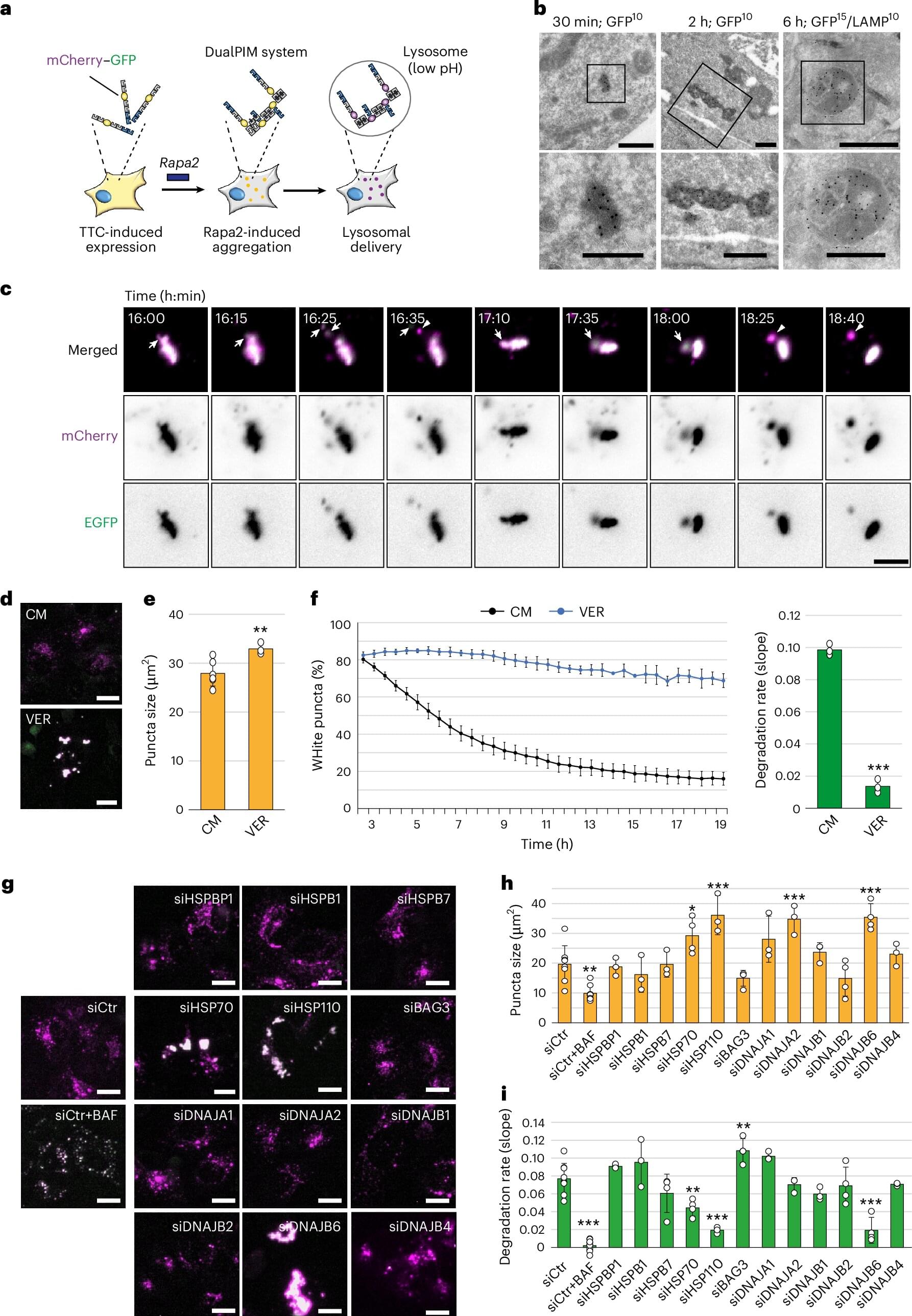
Toggle light / dark theme

Is your heart aging too fast? MRI technique reveals how unhealthy lifestyles can add decades

Scientists at the University of East Anglia (UEA) have developed a new way of uncovering the “true age” of a heart using MRI.

Research accepted for publication European Heart Journal Open shows how an MRI scan can reveal your heart’s functional age—and how unhealthy lifestyles can dramatically accelerate this figure. The paper is titled “Cardiac MRI Markers of Ageing: A Multicentre, Cross-sectional Cohort Study.”

It is hoped that the findings could transform how heart disease is diagnosed—offering a lifeline to millions by catching problems before they become deadly.

Lysosome destabilization found to drive iron-dependent cell death in cancer

The duplication and division of cells is critical to keeping all multicellular organisms alive. But the opposite process is equally important: cell death. Controlled death of cells, or programmed cell death, is also necessary for the proper development and function of the body. It has also been a focus of researchers developing treatments for cancer by finding ways to activate the cell death of cancer cells themselves.

Ferroptosis is a recently discovered form of programmed and has been a promising target for the development of cancer treatments. It is mediated by iron molecules, with the cell dying through the degradation of the phospholipid bilayer by oxidation, a process called . However, recent studies have shown that certain cancer cells are less susceptible to ferroptosis, raising concerns that this resistance could pose a barrier to future therapeutics.

In a paper published in Nature Communications, researchers from Kyushu University, using cultured cells and mice, found that the lipid peroxidation of the lysosomes—the organelle responsible for degrading and recycling molecules in a cell—plays a critical role in the execution of ferroptosis.

Making AI models more trustworthy for high-stakes contexts, like classifying diseases in medical images

Antimicrobial resistance (AMR) presents a serious challenge in today’s world. The use of antimicrobials (AMU) significantly contributes to the emergence and spread of resistant bacteria. Companion animals gain recognition as potential reservoirs and vectors for transmitting resistant microorganisms to both humans and other animals. The full extent of this transmission remains unclear, which is particularly concerning given the substantial and growing number of households with companion animals. This situation highlights critical knowledge gaps in our understanding of risk factors and transmission pathways for AMR transfer between companion animals and humans. Moreover, there’s a significant lack of information regarding AMU in everyday veterinary practices for companion animals. The exploration and development of alternative therapeutic approaches to antimicrobial treatments of companion animals also represents a research priority. To address these pressing issues, this Reprint aims to compile and disseminate crucial additional knowledge. It serves as a platform for relevant research studies and reviews, shedding light on the complex interplay between AMU, AMR, and the role of companion animals in this global health challenge. This Reprint is especially addressed to companion animal veterinary practitioners as well as all researchers working on the field of AMR in both animals and humans, from a One Health perspective.

Predicting relapse of autoimmune small vessel vasculitis

Neutrophils, one of the immune system warriors that were thought to be all the same, turn out to be diverse. Unfortunately, these cells are also active in autoimmune diseases. New research has found that a certain subpopulation of these white blood cells can predict disease relapse at an early stage, which may enable improved personalized treatment.

In a study published in Nature Communications, a multi-institutional research team investigated which cell types dominate the blood of patients at the early stage of anti-neutrophil cytoplasmic antibody (ANCA)-associated vasculitis, which is caused by inflammation in the blood vessels and can disrupt organ function.

“Figuring out the mechanism of this disease, which is poorly understood, will help us understand autoimmune dysregulation in neutrophils. This could aid in the development of new drugs tailored for each patient,” says the lead author of the study. “Because we want to understand the dynamics of neutrophil behavior at the cell level in the early stages of the disease, for this study we recruited new patients that had not yet been treated.”

Inhibition of NADPH Oxidase (NOX) 2 Mitigates Colitis in Mice with Impaired Macrophage AMPK Function

Macrophage adenosine monophosphate-activated protein kinase (AMPK) limits the development of experimental colitis. AMPK activation inhibits NADPH oxidase (NOX) 2 expression, reactive oxygen species (ROS) generation, and pro-inflammatory cytokine secretion in macrophages during inflammation, while increased NOX2 expression is reported in experimental models of colitis and inflammatory bowel disease (IBD) patients. Although there are reductions in AMPK activity in IBD, it remains unclear whether targeted inhibition of NOX2 in the presence of defective AMPK can reduce the severity of colitis. Here, we investigate whether the inhibition of NOX2 ameliorates colitis in mice independent of AMPK activation. Our study identified that VAS2870 (a pan-Nox inhibitor) alleviated dextran sodium sulfate (DSS)-induced colitis in macrophage-specific AMPKβ1-deficient (AMPKβ1LysM) mice.

How morphogens steer early brain development by guiding stem cell gene activity

Just a few weeks after conception, stem cells are already orchestrating the future structure of the human brain. A new Yale-led study shows that, early in development, molecular “traffic cops” known as morphogens regulate the activation of gene programs that initiate stem cells’ differentiation into more specialized brain cells.

The Yale team found that sensitivity to these signaling morphogens can vary not only between stem cells from different donors, but between stem cells derived from the same individual.

“This is a new chapter in understanding how we develop and how development can be influenced by genomic changes between people and by within individuals,” said Flora Vaccarino, the Harris Professor in the Child Study Center at the Yale School of Medicine (YSM) and co-senior author of the research, published in the journal Cell Stem Cell.

FDA-designated orphan drug could increase radiation efficacy in lung cancer

An FDA-designated orphan drug that can target a key vulnerability in lung cancer shows promise in improving the efficacy of radiation treatments in preclinical models, according to a study by UT Southwestern Medical Center researchers. The findings, published in Science Advances, suggest a new way to enhance the response to radiotherapy by inhibiting DNA repair in lung cancer cells.

“This study was motivated by challenges faced by millions of cancer patients undergoing radiation therapy, where treatment-related toxicities limit both curative potential and the patient’s quality of life,” said principal investigator Yuanyuan Zhang, M.D., Ph.D., Assistant Professor of Radiation Oncology and a member of the Harold C. Simmons Comprehensive Cancer Center at UT Southwestern.

Prior research, including from the laboratory of co-investigator Ralph J. DeBerardinis, M.D., Ph.D., Professor and Director of the Eugene McDermott Center for Human Growth and Development, Professor in Children’s Medical Center Research Institute at UT Southwestern, and co-leader of the Cellular Networks in Cancer Research Program in the Simmons Cancer Center, has demonstrated that altered metabolic pathways in allow them to survive, grow, and spread. But the role of metabolism in enhancing radiation efficacy has not been thoroughly explored.

/* */