As CIOs, we must lead the way in diagnosing these cancers early. Here’s how:
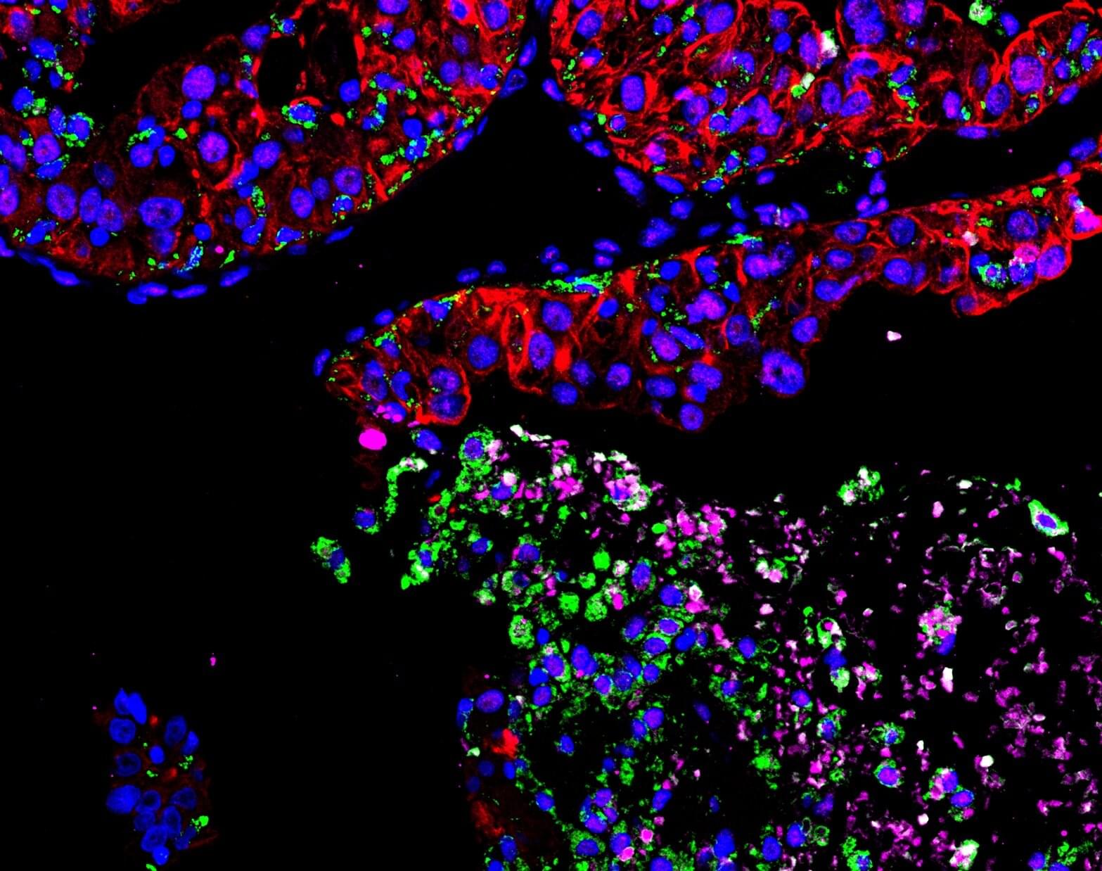
Cancer specialists have long known that anemia, caused by a lack of healthy red blood cells, often arises when cancer metastasizes to the bone, but it’s been unclear why. Now, a research team led by Princeton University researchers Yibin Kang and Yujiao Han has uncovered exactly how this happens in metastatic breast cancer, and it involves a type of cellular hijacking. The research aims to help slow down bone metastasis—one of cancer’s deadliest forms.
In a study published in the journal Cell on September 3, Kang and Han reveal that cancer cells effectively commandeer a specialized cell that normally recycles iron in the bone, known as an erythroblast island (EBI) macrophage. This both deprives red blood cells of necessary iron and helps the tumor continue to grow in the bone.
Understanding metastatic cancer—or cancer that grows and spreads in other parts of the body beyond the original tumor site—is critically important. It is one of the deadliest forms of cancer and there is no cure. Of patients who die from breast and prostate cancer, 70% have bone metastasis.

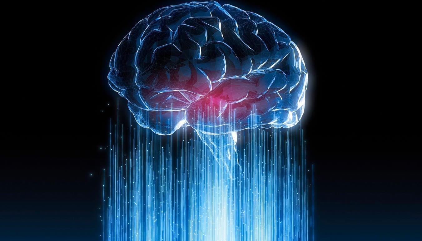
Neural networks are computing systems designed to mimic both the structure and function of the human brain. Caltech researchers have been developing a neural network made out of strands of DNA instead of electronic parts that carries out computation through chemical reactions rather than digital signals.
An important property of any neural network is the ability to learn by taking in information and retaining it for future decisions. Now, researchers in the laboratory of Lulu Qian, professor of bioengineering, have created a DNA-based neural network that can learn. The work represents a first step toward demonstrating more complex learning behaviors in chemical systems.
A paper describing the research appears in the journal Nature on September 3. Kevin Cherry, Ph.D., is the study’s first author.
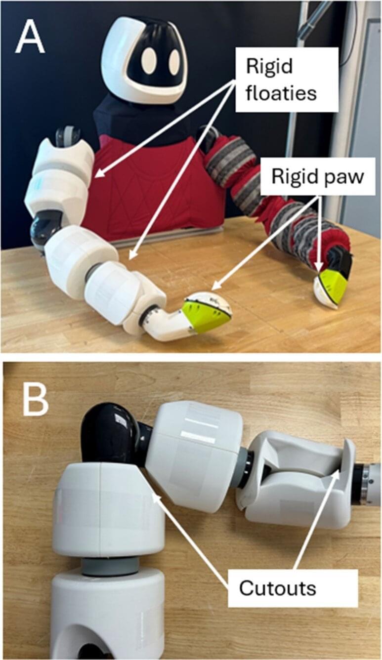
For all their technological brilliance, from navigating distant planets to performing complex surgery, robots still struggle with a few basic human tasks. One of the most significant challenges is dexterity, which refers to the ability to grasp, hold and manipulate objects. Until now, that is. Scientists from the Toyota Research Institute in Massachusetts have trained a robot to use its entire body to handle large objects, much like humans do.
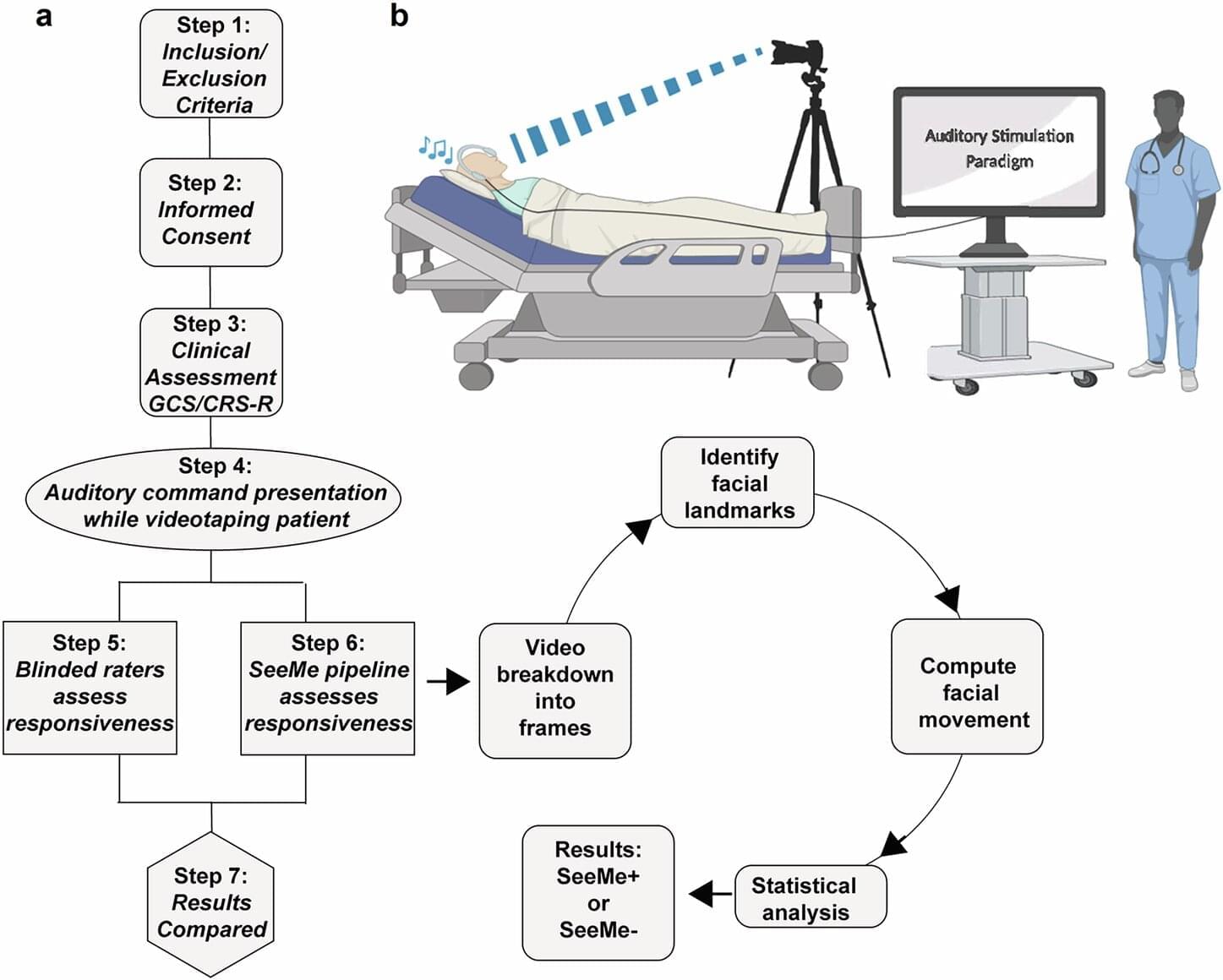
SeeMe, a computer vision tool tested by Stony Brook University researchers, was able to detect low-amplitude, voluntary facial movements in comatose acute brain injury patients days before clinicians could identify overt responses.
Close friends know that I have a standing “do not unplug” order should I ever fall into an unresponsive state. If there is even a flicker of a chance that the mind is still working, I will be fine. Keep me plugged in and hang a “do not disturb” sign on whatever apparatus is keeping me alive.
It’s not like you can know in advance what it’s like, but it seems relaxed enough, with plenty of time to think, and I haven’t really gained anything useful from conversations with other humans in years (aside from my editors who always provide valuable information). If it is at all like sleeping, there might be dreams, so, perchance, that’s what I’d be doing in a comatose state. But for the friends by my bedside, how to be certain that the mind is still flickering?

Researchers at Beijing Genomics and IMDEA Nanociencia institutes have introduced a novel method that could significantly accelerate efficiency and reduce the cost of handling fluidics in DNA sequencing.
Traditional DNA sequencing relies on flow cells, where liquid reagents are repeatedly pumped in and out for each of the sequencing reactions. For large-scale sequencing, this process involves immersing silicon wafers into reagents—a method that works well at industrial scale but is impractical for smaller labs or clinical settings, where sample sizes are limited and drying effects become a problem.
The new approach turns that process on its head. Instead of pumping fluids through a chamber, the researchers use a roll-to-roll technique that gently shears the liquid across the surface. This dramatically improves efficiency, allowing reagents to be replaced more quickly and uses up to 85 times less material. As a result, DNA sequencing that once took days can now be completed in under 12 hours, with significantly lower reagent costs.

Stanford Medicine scientists have developed a brain-computer interface that detects inner speech from speech-impaired patients, in a step toward restoring rapid communication.

A Harvard-affiliated study suggests that daily vitamin D supplementation may help slow biological aging by protecting DNA and preserving telomere length. The VITAL trial, which tracked over 1,000 adults for four years, found that participants taking 2,000 IU of vitamin D daily experienced less telomere shortening, effectively reducing biological aging by nearly three years.