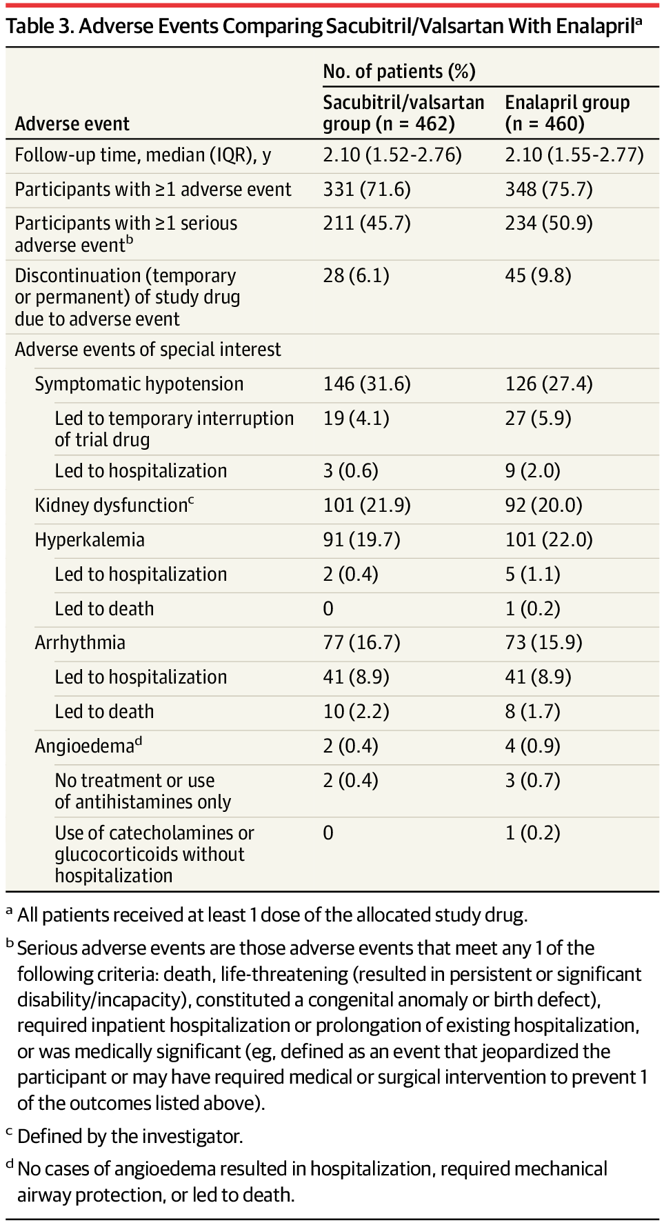
Toggle light / dark theme

Protein engineering research reveals the mysteries of life, enabling advances in pharmaceuticals

Proteins are so much more than nutrients in food. Virtually every reaction in the body that makes life possible involves this large group of molecules. And when things go wrong in our health, proteins are usually part of the problem.

In certain types of heart disease, for instance, the proteins in cardiac tissue, seen with , are visibly disordered. Alex Dunn, professor of chemical engineering, describes proteins like the beams of a house: “We can see that in unhealthy heart muscle cells, all of those beams are out of place.”

Proteins are the workhorses of the cell, making the biochemical processes of life possible. These workhorses include enzymes, which bind to other molecules to speed up reactions, and antibodies that attach to viruses and prevent them from infecting cells.

Intestinal Infections can Alter Bile Composition & Immunity

Researchers have used a mouse model to show that infections in the intestine can change the composition of bile, a fluid that is generated in the liver and is crucial to digestion. Bile aids in the absorption of fat and contributes to defense against infections. This study has suggested that intestinal infections can alter microbes in the gastrointestinal tract, or the microbiome, and modify the immune system. Although the work was conducted in mice, the researchers suggested that their conclusions also apply to humans. The findings have been reported in Nature Microbiology.

“The changes we detected in the composition of bile with infection are beneficial for the intestine to clear infection,” said corresponding study author Matthew Waldor, MD, PhD, of Brigham and Women’s Hospital. “Our findings reveal the intricate and dynamic nature of bile composition, shedding new light on the liver’s critical role in defending the intestine from infection. These insights enhance our understanding of the liver’s broader functions in regulating physiological stability and metabolic processes.”

Study identifies ‘turncoat’ cells that fight—and aid—cancer

Yale researchers have made an unexpected discovery—turncoat T cells that help a tumor evade other cancer-fighting immune T cells—in a study of patients living with advanced melanoma.

The study by Yale Cancer Center (YCC) researchers at Yale School of Medicine (YSM) discovered that not all CD8+ T cells are allies in a body’s fight against . Patients living with severe who had increased levels of suppressor, regulatory CD8+ T cells had worse survival outcomes.

The study is published in the journal Nature Immunology.

How Is Cell Death Essential to Life?

Death might seem like a pure loss, the disappearance of what makes a living thing distinct from everything else on our planet. But zoom in closer, to the cellular level, and it takes on a different, more nuanced meaning. There is a challenge in simply defining what makes an individual cell alive or dead. Scientists today are working to understand the various ways and reasons that cells disappear, and what these processes mean to biological systems. In this episode, cellular biologist Shai Shaham talks to Steven Strogatz about the different forms of cell death, their roles in evolution and disease, and why the right kinds and patterns of cell death are essential to our development and well-being.

Listen on Apple Podcasts, Spotify, TuneIn or your favorite podcasting app, or you can stream it from Quanta.

Prevention and screening outpace treatment advances for averting death from five cancer types, study reveals

Improvements in cancer prevention and screening have averted more deaths from five cancer types combined over the past 45 years than treatment advances, according to a modeling study led by researchers at the National Institutes of Health (NIH).

The study, published Dec. 5, 2024, in JAMA Oncology, looked at deaths from breast, cervical, colorectal, lung, and prostate cancer that were averted by the combination of prevention, , and advances.

The researchers focused on these five cancers because they are among the most common causes of cancer deaths and strategies exist for their prevention, early detection, and/or treatment. In recent years, these five cancers have made up nearly half of all new cancer diagnoses and deaths.

Algorithm analyzes multiple mammograms to improve breast cancer risk prediction

A new study from Washington University School of Medicine in St. Louis describes an innovative method of analyzing mammograms that significantly improves the accuracy of predicting the risk of breast cancer development over the following five years.

Using up to three years of previous mammograms, the new method identified individuals at high risk of developing 2.3 times more accurately than the standard method, which is based on questionnaires assessing clinical risk factors alone, such as age, race and family history of breast cancer.

The study is published Dec. 5 in JCO Clinical Cancer Informatics.

Alignment of Cells Affects Secondary Tumor Growth

Cell–cell alignment and a background of stationary cells together shape the emergence of cellular clusters in a primary tumor.

In a cancer patient, tumor cells that circulate throughout the body in clusters pose a greater threat of metastasis than those that circulate individually. Those clusters are thought to come together while the cells are still within the primary tumor, but researchers still don’t understand the formation mechanism. Quirine Braat at Eindhoven University of Technology in the Netherlands and her colleagues have now used computer simulations to identify some of the factors at play [1].

The team used a computational lattice model of cells and tissues (the cellular Potts model) to examine a 2D layer of two types of cells—one motile (able to move) and one nonmotile. The tendency of the motile cells to migrate was represented in the model by an external force applied to each one. For a given cell, this force could align strongly or weakly with the forces acting on its neighboring cells.

Polarization photodetector that mimics desert ant offers pathway for more sensitive, miniaturized imaging systems

Polarization photodetectors (pol-PDs) have widespread applications in geological remote sensing, machine vision, and biological medicine. However, commercial pol-PDs usually require bulky and complicated optical components and are difficult to miniaturize and integrate.

Chinese researchers have made important progress in this area by developing an on-chip integrated polarization .

This study, published in Science Advances on Dec. 4, was conducted by Prof. Li Mingzhu’s group from the Technical Institute of Physics and Chemistry of the Chinese Academy of Sciences.

Bioprinting technique creates functional tissue 10x faster

Three-dimensional (3D) printing isn’t just a way to produce material products quickly. It also offers researchers a way to develop replicas of human tissue that could be used to improve human health, such as building organs for transplantation, studying disease progression and screening new drugs. While researchers have made progress over the years, the field has been hampered by limited existing technologies unable to print tissues with high cell density at scale.

A team of researchers from Penn State have developed a novel bioprinting technique that uses spheroids, which are clusters of cells, to create complex tissue. This new technique improves the precision and scalability of tissue fabrication, producing tissue 10-times faster than existing methods. It further opens the door to developing functional tissues and organs and progress in the field of regenerative medicine, the researchers said.

They published their findings in Nature Communications.