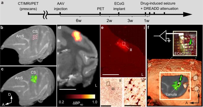
Pharmacological and surgical treatments of epilepsy can have unsatisfactory outcomes, so a more targeted and on-demand approach is desirable. Here, the authors demonstrate the usage of inhibitory chemogenetics in male nonhuman primates to attenuate the magnitude and spread of cortical seizures and subsequent body convulsions.
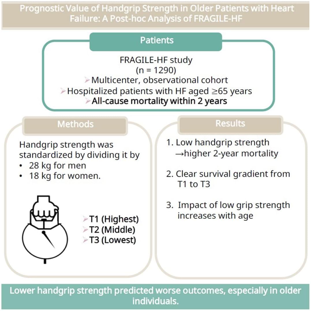
Lower handgrip strength is associated with higher 2-year death risk in older heart failure patients, with stronger impact at advanced ages. @NakadeTaisuke@yuya_matsue
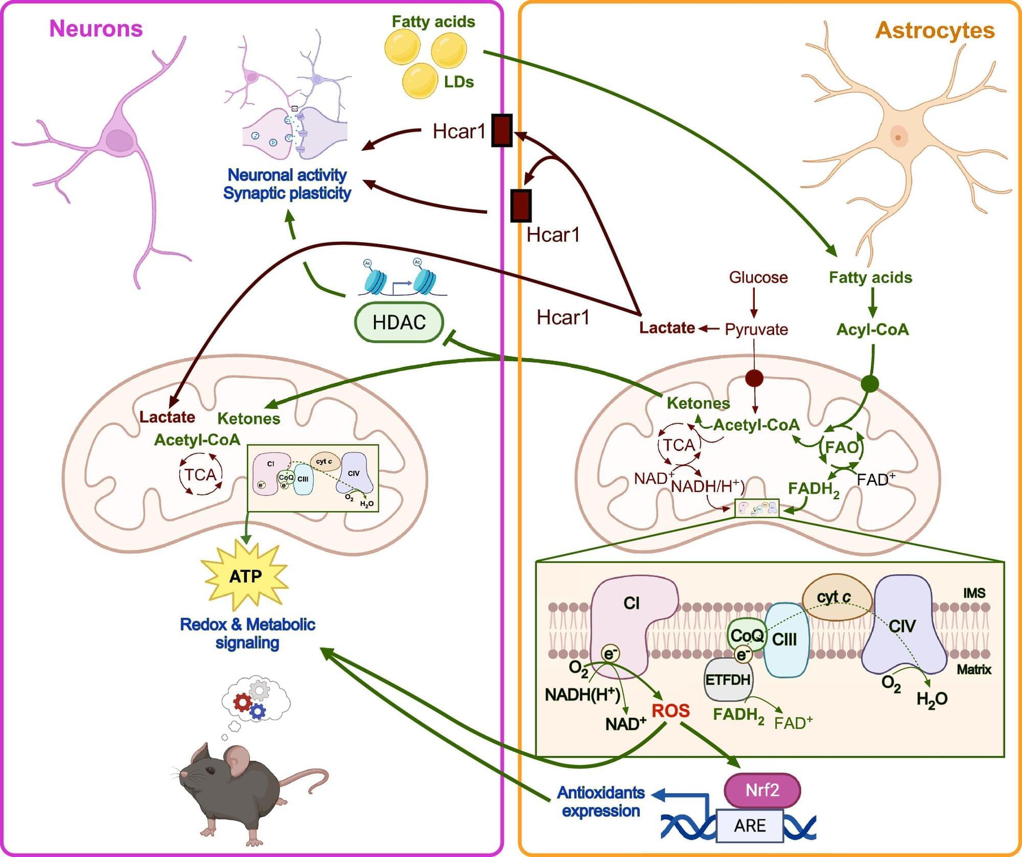
Glia previously thought to be support cells of brain but recent evidence suggest that the astrocytes, the most abundant glial cell type in addition to supplying neurons with lactate via glycolysis also actively engage in lipid metabolism, especially mitochondrial fatty acid β-oxidation.
Researchers in this review integrate astrocytic fatty acid ß-oxidation and ketogenesis, alongside other metabolic pathways converging on reactive oxygen species dynamics, including cholesterol metabolism and peroxisomal β-oxidation.
Astrocytes, the most abundant glial cell type in the central nervous system, have traditionally been viewed from the perspective of metabolic support, particularly supplying neurons with lactate via glycolysis. This view has focused heavily on glucose metabolism as the primary mode of sustaining neuronal function. However, recent research challenges this paradigm by positioning astrocytes as dynamic metabolic hubs that actively engage in lipid metabolism, especially mitochondrial fatty acid β-oxidation. Far from serving solely as an energy source, fatty acid ß-oxidation in astrocytes orchestrates reactive oxygen species-mediated signaling pathways that modulate neuron-glia communication and cognitive outcomes.
The first NMNH human trial shows NAD+ levels increased up to 3x in 90 days. Here’s what the data actually reveal—and what’s still missing. Some links are affiliate links so we will earn a commission when they are used to purchase products.
The first human clinical trial results for NMNH are here. In this 90-day randomized, double-blind, placebo-controlled study, NMNH increased NAD+ levels up to 3x in healthy adults, with participants reporting improvements in energy and fatigue. But before you get too excited, there are important limitations to understand. In this video, I break down the trial design, explain what the NAD+ increases actually mean, review the subjective outcomes like energy and emotional well-being (measured via SF-36), and discuss why the biological age claims are difficult to interpret. I also cover safety data and what we still need to know. This is promising early data for NMNH vs NMN, but it’s unpublished, lacks key details, and needs independent replication. Here’s everything you need to know about what this trial does and doesn’t tell us. Key topics: NMNH clinical trial, NAD+ boosters, NMN vs NMNH, longevity supplements, anti-aging research, NAD+ levels, UthPeak study, Phase I trial results.
📚 Chapters. 0:00 — Introduction & Trial Overview What this video covers and the trial basics. 1:39 — Trial Design & Methodology Study structure, participants, and objectives. 2:31 — NAD+ Results The primary outcome: dose-dependent increases. 3:18 — Subjective Outcomes & Limitations Energy, mood, biological age claims, and why interpretation is difficult. 6:26 — Safety & Final Thoughts Tolerability data and what comes next.
🌐Links in this video. NMNH Clinical Trial https://www.clinicaltrials.gov/study/.… nicotinamide mononucleotide is a new and potent NAD+ precursor in mammalian cells and mice https://faseb.onlinelibrary.wiley.com… Reduced Nicotinamide Mononucleotide (NMNH) Potently Enhances NAD+ and Suppresses Glycolysis, the TCA Cycle, and Cell Growth https://pubmed.ncbi.nlm.nih.gov/33793… *************************************** Health claims Disclosure: Information provided on this video is not a substitute for direct, individual medical treatment or advice. Please consult with your doctor first. Products or services mentioned in this video are not a recommendation. Audio Copyright Disclaimer Please note that we have full authorization to the music that we used in our videos as they were created using the service WeVideo which provides the rights to the music. The rights are detailed in the terms of use that can be reviewed here https://www.wevideo.com/terms-of-use and any following inquiries should be addressed to [email protected]. ********************************************** #nmnh #nad #nmn. Reduced nicotinamide mononucleotide is a new and potent NAD+ precursor in mammalian cells and mice. https://faseb.onlinelibrary.wiley.com… Reduced Nicotinamide Mononucleotide (NMNH) Potently Enhances NAD+ and Suppresses Glycolysis, the TCA Cycle, and Cell Growth. https://pubmed.ncbi.nlm.nih.gov/33793…
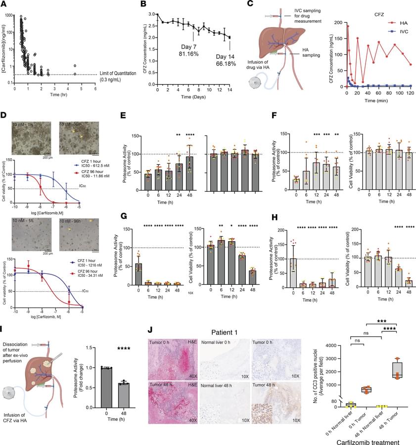
A rebrand for proteasome inhibition in solid tumors.
In this Research Letter, Jonathan M. Hernandez & team investigate the potential of hepatic artery infusion pump delivery of carfilzomib, to continuously direct a large dose to the tumor with least hepatic toxicity.
Address correspondence to: Jonathan M. Hernandez, National Cancer Institute, NIH, 10 Center Drive, Room 4W-3752, Bethesda, Maryland, 20,892, USA. Email: Jonathan. [email protected].
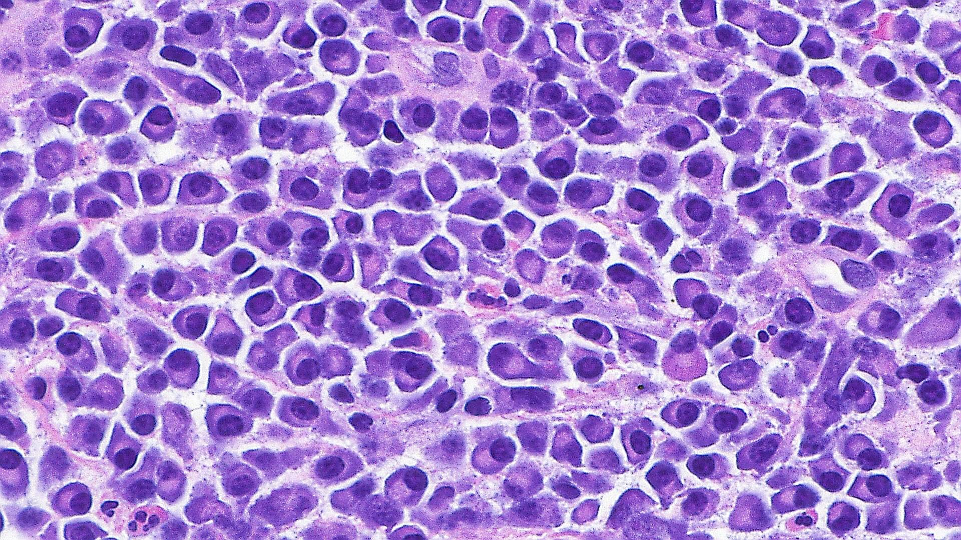
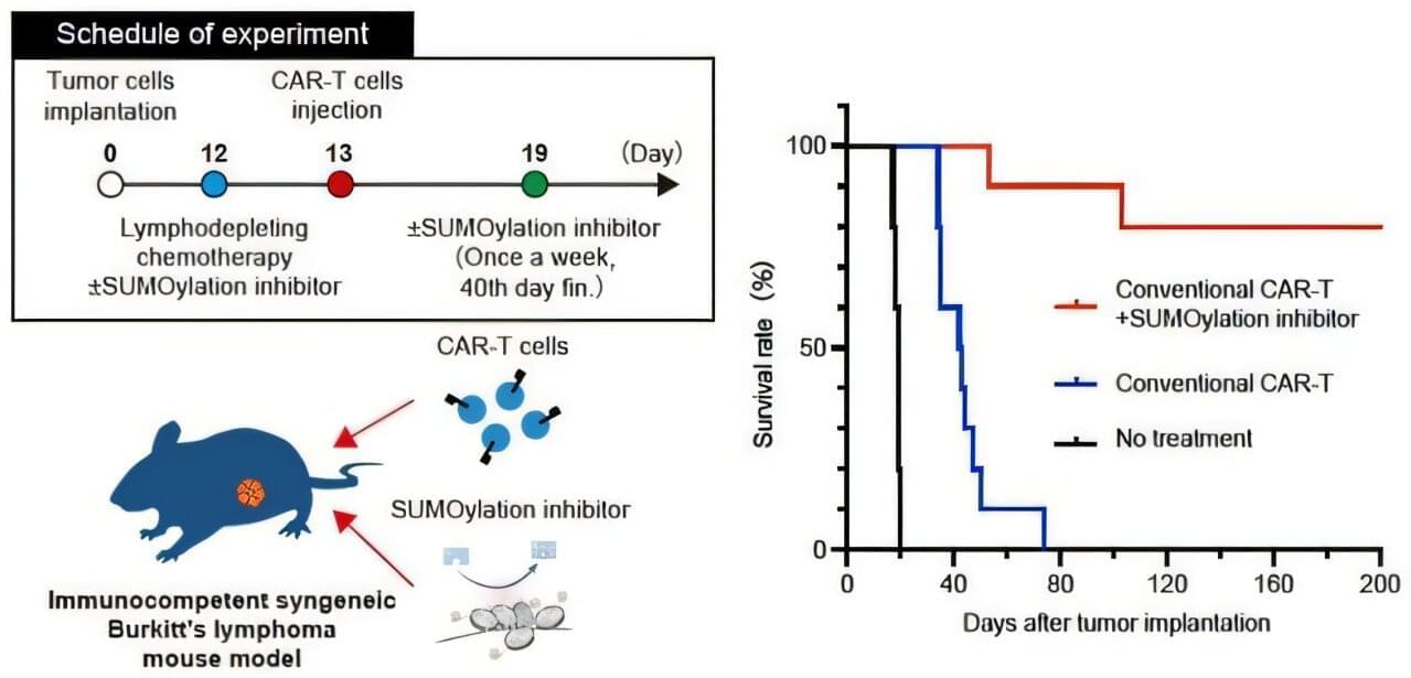
Burkitt’s lymphoma is a rare and aggressive blood cancer characterized by a translocation of the MYC gene. It occurs most often in children and young adults. In recent years, CAR-T cell therapy—often referred to as a “living drug” and administered as a single dose—has been approved for certain types of blood cancer, offering hope for a cure even in severe cases. However, its effectiveness against Burkitt’s lymphoma has been limited. Moreover, developing drugs that directly target MYC—the root cause of this cancer—has proven challenging for decades.
Recently, a study led by Dr. Hiroshi Kotani, Assistant Professor at Kanazawa University in collaboration with a scientist at Roswell Park Comprehensive Cancer Center in Buffalo, NY, U.S., revealed that a SUMOylation inhibitor can suppress MYC activity. Building on this finding, the research team investigated whether combining CAR-T therapy with the SUMOylation inhibitor TAK-981 could improve outcomes for Burkitt’s lymphoma. The research is published in Signal Transduction and Targeted Therapy.
The team first confirmed that the SUMOylation inhibitor effectively slowed the growth of Burkitt’s lymphoma cells and altered their signaling pathways. They then examined the inhibitor’s effect on CAR-T cells and discovered a dual role: While it initially activated the CAR-T cells in a way that could hinder long-term effectiveness, it also triggered a built-in “safety brake” mechanism. These insights suggested that using only a limited dose of the inhibitor could maximize the benefits of CAR-T therapy as a durable, living treatment.
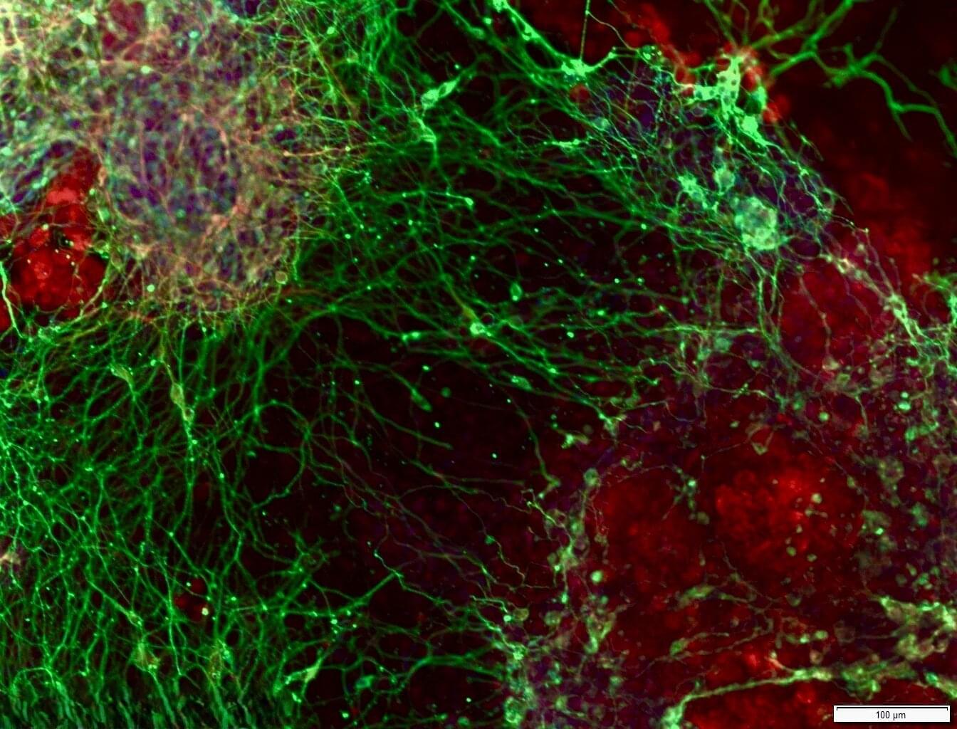
Which genes are required for turning embryonic stem cells into brain cells, and what happens when this process goes wrong? In a new study published today in Nature Neuroscience, researchers led by Prof. Sagiv Shifman from The Institute of Life Sciences at The Hebrew University of Jerusalem, in collaboration with Prof. Binnaz Yalcin from INSERM, France, used genome-wide CRISPR knockout screens to identify genes that are needed for early brain development.
The study set out to answer a straightforward question: which genes are required for the proper development of brain cells?
Using CRISPR-based gene-editing methods, the researchers systematically and individually “switched off” roughly 20,000 genes to study their role in brain development. They performed the screen in embryonic stem cells while the cells changed into brain cells. By disrupting genes one by one, the team could see which genes are required for this transition to proceed normally.
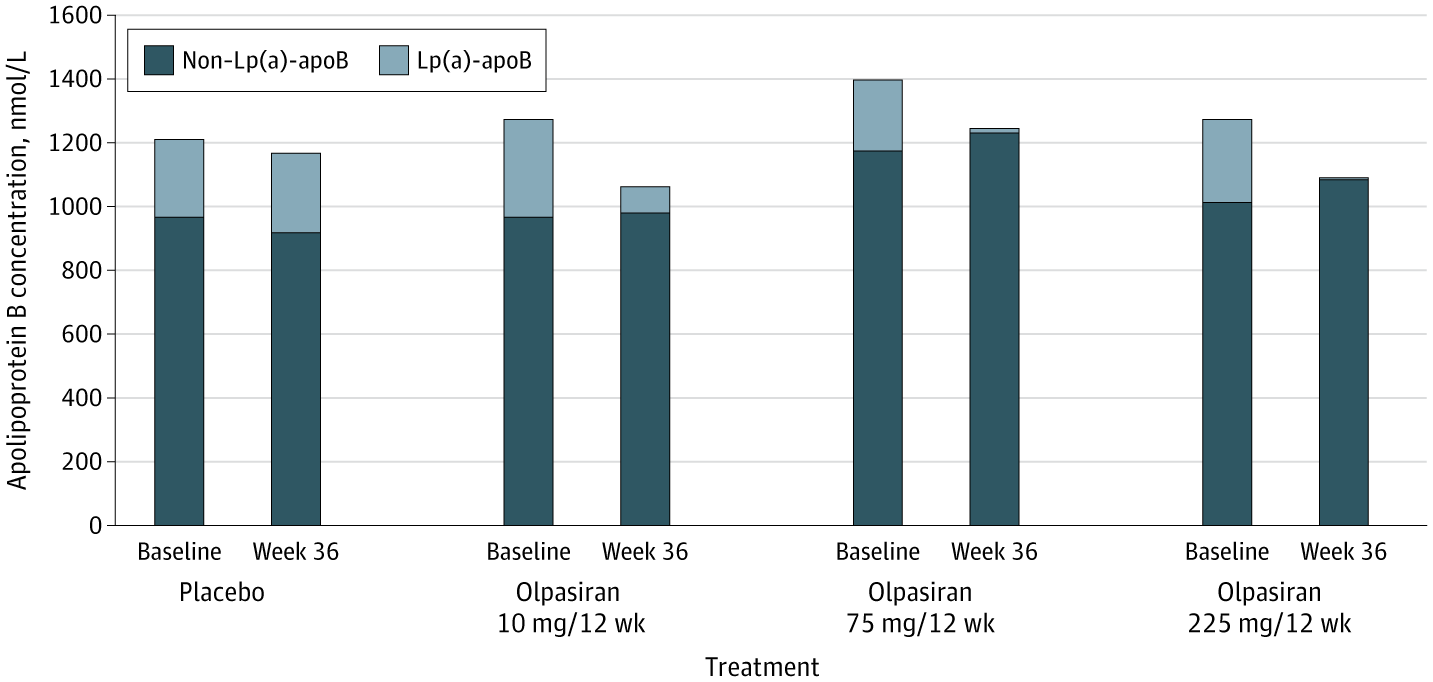
Small-interfering RNA olpasiran reduced lipoprotein(a)–apolipoprotein B particles by 95% with minimal rise in non–Lp(a)-apoB, lowering total apoB concentration in patients with cardiovascular disease.
This secondary analysis of the OCEAN(a)-DOSE randomized clinical trial investigates the effect of the small-interfering RNA olpasiran on atherogenic lipoproteins.
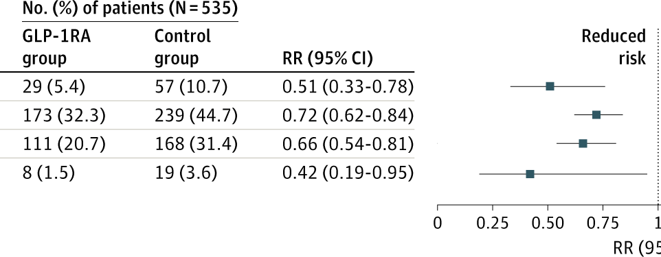
This retrospective cohort study reported an association between GLP1RA use and a lower risk of acute asthma exacerbations in adolescents with overweight or obesity, suggesting a potential dual benefit for this population.
This cohort study investigates the association between glucagonlike peptide-1 receptor agonist use and the risk of acute asthma exacerbations among adolescents with overweight or obesity and asthma.