Despite advances in the management of advanced clear cell renal cell carcinoma (ccRCC), robust biomarkers for prognosis and therapeutic response prediction remain elusive.
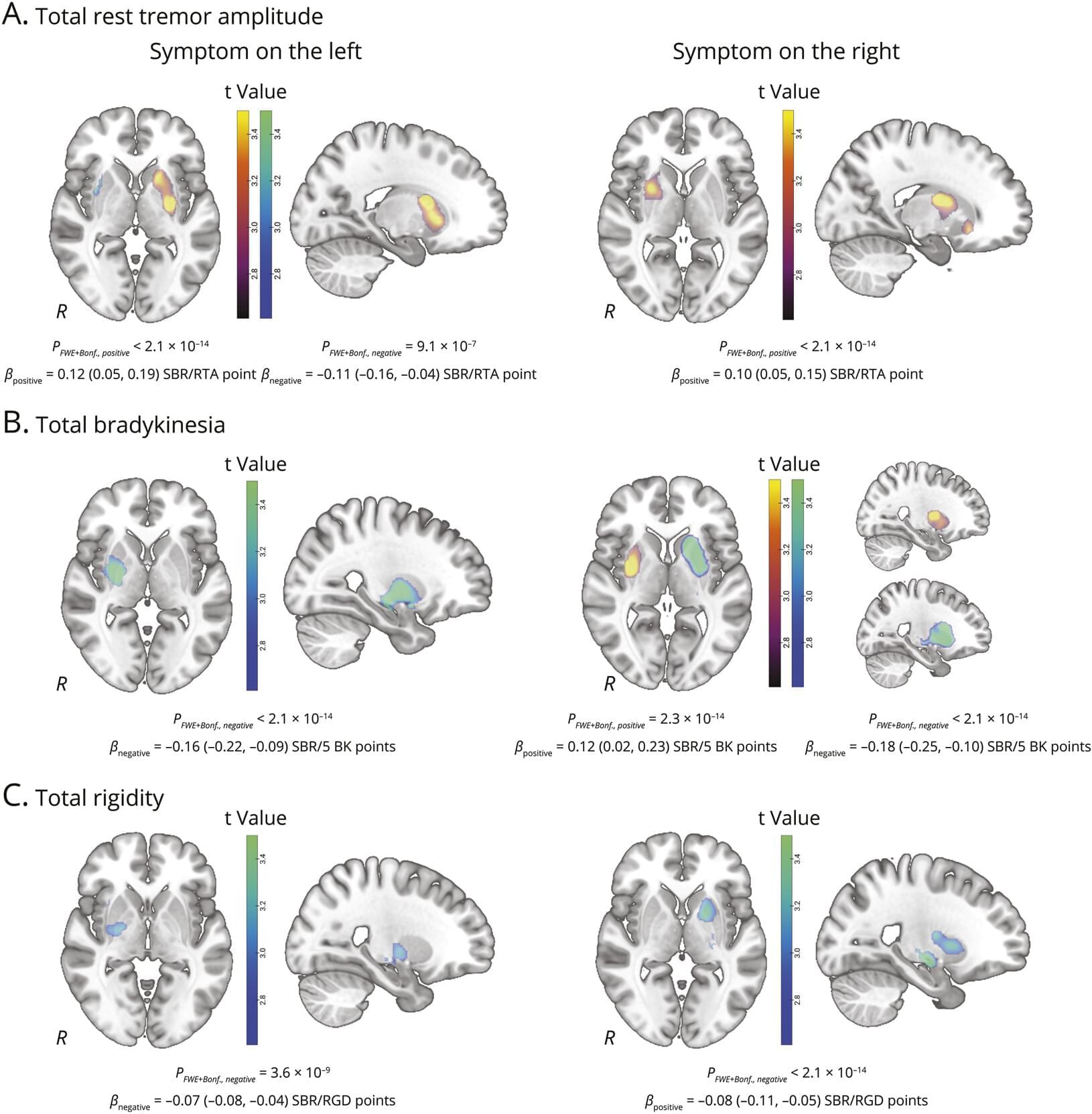
Sickle cell disease is often thought of solely as a blood disorder, but new research from the Wood Neuro Research Group provides measurable evidence that it can reshape how brain networks function. Previous neuroimaging studies have relied on functional connectivity to show that adults with sickle cell disease may experience changes in how brain networks communicate among one another, potentially compensating for reduced oxygen delivery. However, this method is limited in determining the directionality or influence between networks.
“Red blood cells that carry oxygen to the brain are altered by the disease, resulting in reduced oxygen delivery to all regions of the brain and long-term changes in how it functions,” outlined Nahom Mossazghi, biomedical engineering Ph.D. student and the study’s first author. “The brain actively recruits other regions to help process information, which we do not see in people without the disease.”
The study, published in Human Brain Mapping, used MRI and advanced analytical tools originally developed in economics to examine how different brain networks influence one another. Instead of functional connectivity, effective connectivity was used to address a gap in the field and interpret how specific networks support one another in response to the disease-related changes.
Many patients suffer from epilepsy that cannot be controlled by current medications. Surgical removal of epileptogenic brain regions is effective in only about half of cases, and not all patients are eligible for the procedure. For these individuals, therapeutic options remain severely limited. Researchers from the Paris Brain Institute and the Institut du Fer à Moulin in Paris have now taken an important step forward: they have identified two molecules capable of reducing seizure frequency by targeting a mechanism that has so far received little attention. Their findings are published in Proceedings of the National Academy of Sciences.
For the brain to function normally, it must continuously regulate its electrical activity. One of the key mechanisms involved is GABAergic signaling, a natural inhibitory system that controls neuronal activity and prevents the electrical bursts that characterize epileptic seizures. This braking system depends on a delicate balance: the concentration of chloride inside neurons.
An ion transporter known as KCC2 is responsible for removing excess chloride from nerve cells. When it functions poorly—as observed in many neurological disorders, including mesial temporal lobe epilepsy, the most common form of focal epilepsy in adults—chloride accumulates inside neurons. As a result, GABAergic signals, instead of inhibiting neuronal activity, can paradoxically excite it.
EPFL researchers have developed a light-based method that can produce proteins that switch states, respond to signals, and even compute, using light and the cell cycle.
Evolution is biology’s powerful method of engineering. It works by generating many variants of DNA, RNA, and proteins inside cells and letting nature “select” the organism that performs best. Early farmers started taking advantage of evolution by interfering with natural selection and letting only the most productive livestock and crops mate.
In laboratories, researchers have developed methods for directed evolution of proteins, especially enzymes and antibodies, that are used in household detergents, medicine, and industry.
Sherlock et al. examine an IRES RNA that initiates translation of a small downstream coding region within a viral genome. The structure, function, and mechanism of this IRES are interrogated experimentally. Differential translation efficiencies between two IRESs within one viral genome exemplify RNA-structure-based tuning of gene expression.
This RedJournal article presents a first step towards continuous dosimetry in targeted radiopharmaceutical therapy by developing a sparse sensor system to reconstruct continuous time-activity curves in preclinical 177Lu-based therapies, demonstrating high accuracy with short scan times.
177Lu-based radiopharmaceutical therapy (RPT) has shown increasing promise in the treatment of neuroendocrine and metastatic prostate cancer. Delivering optimal radiation dose to tumors while minimizing dose to organs-at-risk (OAR) remains an unmet need due to significant patient-to-patient heterogeneity in treatment response, necessitating multiple snapshots of the in vivo activity distribution. Towards this goal, here we present a high temporal-resolution activity reconstruction method demonstrated on preclinical prostate cancer models.
Editor’s Note: Associate Editor Pablo Penaloza-MacMaster provides context for Long et al. on targeting the host factor HGS-viral membrane protein interaction: https://doi.org/10.1172/JCI200225
While current antivirals primarily target viral proteins, host-directed strategies remain underexplored. Here, we performed a genome-wide CRISPR inhibition (CRISPRi) screening to identify the host protein, hepatocyte growth factor-regulated tyrosine kinase substrate (HGS), facilitating the pan-coronavirus infection both in vitro and in vivo. Mechanistically, HGS interacts with the viral membrane (M) protein, facilitating its trafficking to the ER-Golgi intermediate compartment for virion assembly. Conversely, HGS deficiency caused M retention in the ER, blocking assembly. Leveraging this interaction, we designed M-derived peptides and screened over 5,000 FDA-approved or commonly used drugs, identifying riboflavin tetrabutyrate (RTB).
Researchers at Washington University in St. Louis have developed a novel cell therapy for Alzheimer’s disease using genetically modified astrocytes — the brain’s most abundant cells. By equipping these cells with a chimeric antigen receptor (CAR), scientists enabled them to specifically target and clear beta-amyloid plaques, the toxic protein deposits that accumulate in brain tissue and drive neurodegeneration. In mouse trials, a single injection prevented plaque formation in young healthy rodents and reduced existing plaque levels by half in older mice. While the approach is still being refined to minimize side effects and must be evaluated for human safety, it holds promise both as a preventive measure and as a treatment at various stages of Alzheimer’s. The same technology may eventually be adapted for cancer therapy by reprogramming the cells to target tumor markers.
Alzheimer’s disease (AD) is the leading cause of dementia and is characterized by progressive amyloid accumulation followed by tau-mediated neurodegeneration. Despite advances in anti-amyloid immunotherapies, important limitations remain, highlighting the need for new therapeutic strategies. Here, we introduce anti-amyloid chimeric antigen receptors expressed in astrocytes (CAR-A) and validate their function in vitro. We show that two CAR-A designs reduce amyloid and associated pathology after plaque formation and prevent early plaque deposition in vivo. Single-nucleus RNA sequencing shows that CAR-A treatment induces a distinct glial response to amyloid pathology involving coordinated activity of astrocytes and microglia. Each construct additionally elicits distinctive, receptor-specific effects in astrocytes or microglia.
As metals fuse and sparks fly, one researcher’s work takes the form of DNA, a bacteriophage, a nucleosome, and more.