Baricitinib is an oral selective Janus kinase 1/2 inhibitor with known anti-inflammatory properties. This study evaluates the efficacy and safety of baricitinib in combination with standard of care for the treatment of hospitalised adults with COVID-19.
In this phase 3 double-blind, randomised, placebo-controlled trial, participants were enrolled from 101 centres across 12 countries in Asia, Europe, North America, and South America. Hospitalised adults with COVID-19 receiving standard of care were randomly assigned (1:1) to receive once-daily baricitinib (4 mg) or matched placebo for up to 14 days. Standard of care included systemic corticosteroids, such as dexamethasone, and antivirals, including remdesivir. The composite primary endpoint was the proportion who progressed to high-flow oxygen, non-invasive ventilation, invasive mechanical ventilation, or death by day 28 assessed in the intention-to-treat population. All-cause mortality by day 28 was a key secondary endpoint, and all-cause mortality by day 60 was an exploratory endpoint; both were assessed in the intention-to-treat population. Safety analyses were done in the safety population defined as all randomly allocated participants who received at least one dose of study drug and who were not lost to follow-up before the first post-baseline visit. This study is registered with ClinicalTrials-gov, NCT04421027.
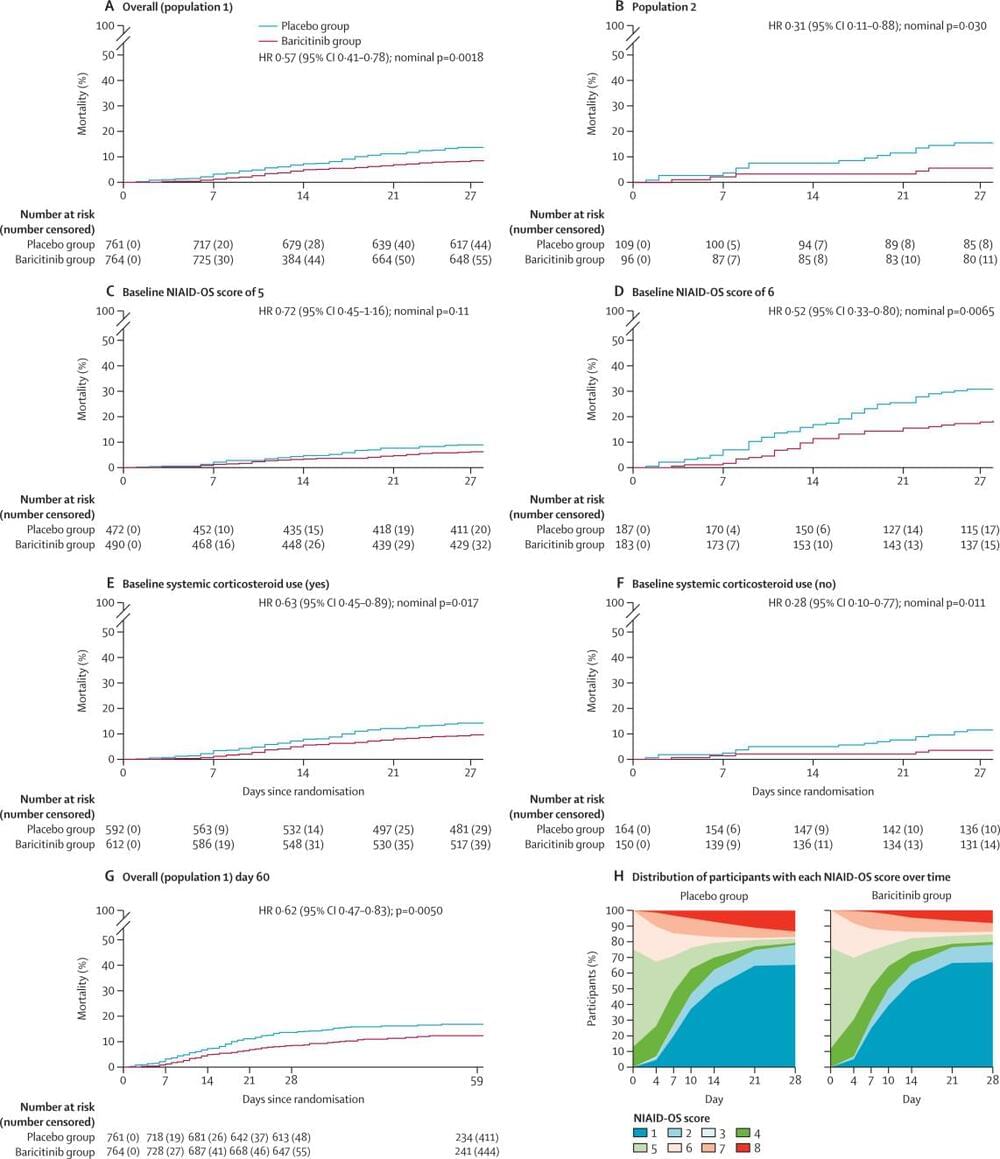
Although there was no significant reduction in the frequency of disease progression overall, treatment with baricitinib in addition to standard of care (including dexamethasone) had a similar safety profile to that of standard of care alone, and was associated with reduced mortality in hospitalised adults with COVID-19.
Eli Lilly and Company.
For the French, Japanese, Portuguese, Russian and Spanish translations of the abstract see Supplementary Materials section.


