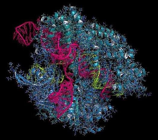
A protein used by viruses could be used as an off switch to make the CRISPR gene editing system safer, researchers say.
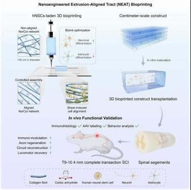
Gu et al. present NEAT, a nanoengineered extrusion-aligned tract bioprinting strategy that fabricates aligned, human neural stem cell-laden collagen hydrogel constructs through shear-induced fibrillar organization. In a rat model of complete spinal cord transection, NEAT enables axonal reconnection and functional locomotor recovery, demonstrating its translational potential for spinal cord repair and neural tissue engineering.
What does it take to turn bold ideas into life-saving medicine?
In this episode of The Big Question, we sit down with @MIT’s Dr. Robert Langer, one of the founding figures of bioengineering and among the most cited scientists in the world, to explore how engineering has reshaped modern healthcare. From early failures and rejected grants to breakthroughs that changed medicine, Langer reflects on a career built around persistence and problem-solving. His work helped lay the foundation for technologies that deliver large biological molecules, like proteins and RNA, into the body, a challenge once thought impossible. Those advances now underpin everything from targeted cancer therapies to the mRNA vaccines that transformed the COVID-19 response.
The conversation looks forward as well as back, diving into the future of medicine through engineered solutions such as artificial skin for burn victims, FDA-approved synthetic blood vessels, and organs-on-chips that mimic human biology to speed up drug testing while reducing reliance on animal models. Langer explains how nanoparticles safely carry genetic instructions into cells, how mRNA vaccines train the immune system without altering DNA, and why engineering delivery, getting the right treatment to the right place in the body, remains one of medicine’s biggest challenges. From personalized cancer vaccines to tissue engineering and rapid drug development, this episode reveals how science, persistence, and engineering come together to push the boundaries of what medicine can do next.
#Science #Medicine #Biotech #Health #LifeSciences.
Chapters:
00:00 Engineering the Future of Medicine.
01:55 Failure, Persistence, and Scientific Breakthroughs.
05:30 From Chemical Engineering to Patient Care.
08:40 Solving the Drug Delivery Problem.
11:20 Delivering Proteins, RNA, and DNA
14:10 The Origins of mRNA Technology.
17:30 How mRNA Vaccines Work.
20:40 Speed and Scale in Vaccine Development.
23:30 What mRNA Makes Possible Next.
26:10 Trust, Misinformation, and Vaccine Science.
28:50 Engineering Tissues and Organs.
31:20 Artificial Skin and Synthetic Blood Vessels.
33:40 Organs on Chips and Drug Testing.
36:10 Why Science Always Moves Forward.
The Big Question with the Museum of Science:
How Elon plans to launch a terawatt of GPUs into space.
## Elon Musk plans to launch a massive computing power of 1 terawatt of GPUs into space to advance AI, robotics, and make humanity multi-planetary, while ensuring responsible use and production. ## ## Questions to inspire discussion.
Space-Based AI Infrastructure.
Q: When will space-based data centers become economically superior to Earth-based ones? A: Space data centers will be the most economically compelling option in 30–36 months due to 5x more effective solar power (no batteries needed) and regulatory advantages in scaling compared to Earth.
☀️ Q: How much cheaper is space solar compared to ground solar? A: Space solar is 10x cheaper than ground solar because it requires no batteries and is 5x more effective, while Earth scaling faces tariffs and land/permit issues.
Q: What solar production capacity are SpaceX and Tesla planning? A: SpaceX and Tesla plan to produce 100 GW/year of solar cells for space, manufacturing from raw materials to finished cells in-house.
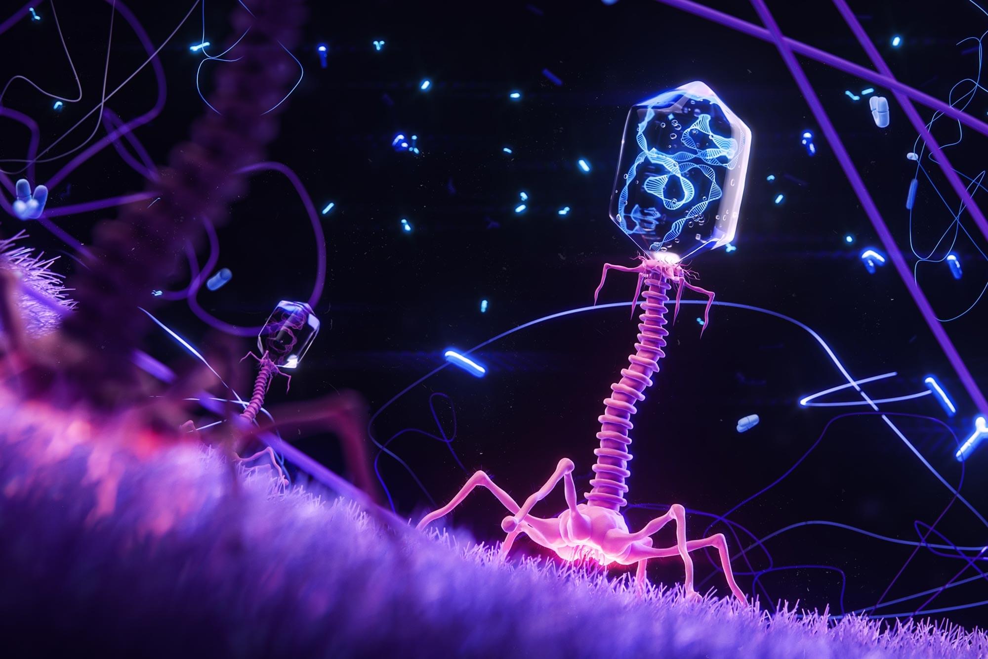
Scientists can now design bacteria-killing viruses from DNA, opening a faster path to fighting superbugs.
Bacteriophages have been used as treatments for bacterial infections for more than a century. Interest in these viruses is rising again as antibiotic-resistant infections become an increasing threat to public health. Even so, progress in the field has been slow. Most research has relied on naturally occurring phages because traditional engineering methods are time consuming and difficult, limiting the development of customized therapeutic viruses.
A fully synthetic phage engineering breakthrough.
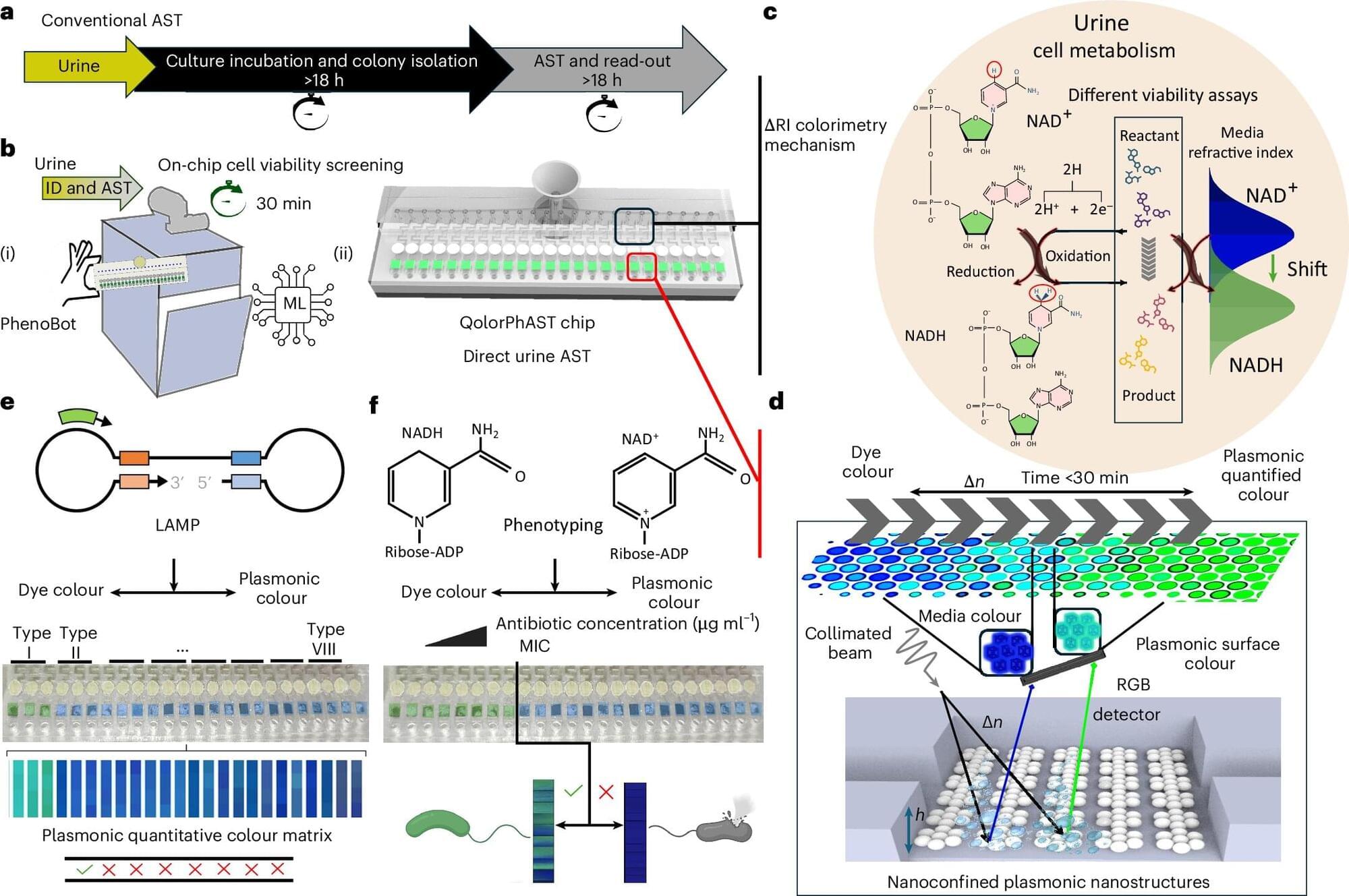
McGill researchers have developed a diagnostic system capable of identifying bacteria—and determining which antibiotics can stop them—in just 36 minutes, a major advance in the global effort to curb antimicrobial resistance (AMR). Current clinical testing methods typically take 48 to 72 hours, leaving physicians without timely guidance.
The researchers say this innovation arrives at a critical moment due to the urgency of the AMR crisis, which arises from bacteria developing resistance to antibiotics.
“We are losing the race against antimicrobial resistance,” said Sara Mahshid, associate professor in the Department of Bioengineering and lead author on the Nature Nanotechnology study. “Every year, more than one million people die, more than from HIV/AIDS or malaria, and delayed treatment is a major driver. Rapid testing isn’t a luxury; it’s the missing link between diagnosis and survival.”
‘Zombie’ coronavirus fragments not only help drive inflammation in long-COVID, but also destroy our immune cells.
A recent study by an international team of more than 30 authors reveals how the destruction of the virus within our body leaves dangerous protein fragments that target specific immune cells, which may explain some of the debilitating consequences millions of people with long-COVID now face.
“These fragments target a specific kind of curvature on the membranes of cells,” explains bioengineer Gerard Wong from the University of California, Los Angeles. “Cells that are spiky, that are star-shaped, or that have lots of tentacles end up getting preferentially suppressed.”
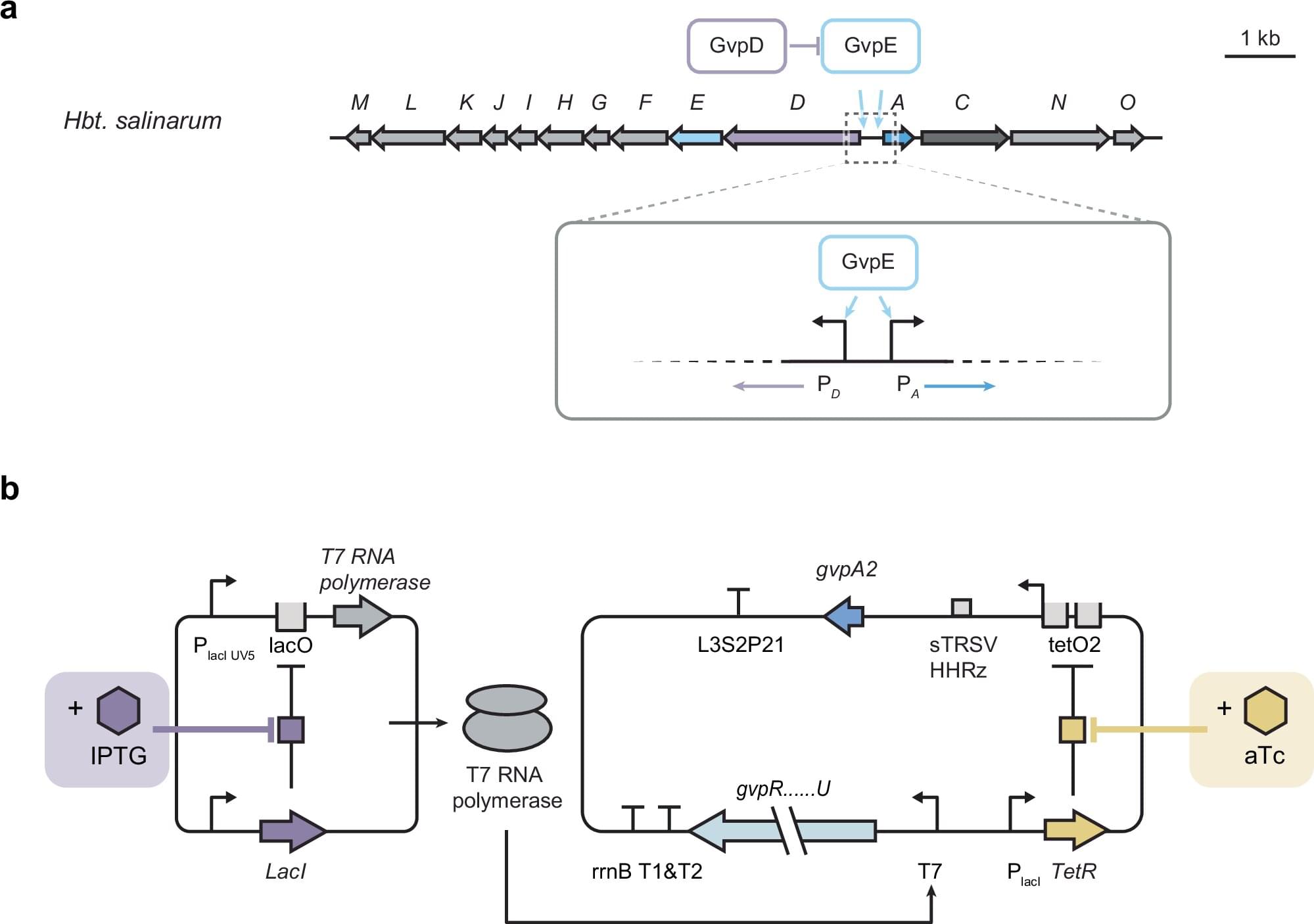
Gas vesicles are among the largest known protein nanostructures produced and assembled inside microbial cells. These hollow, air-filled cylindrical nanostructures found in certain aquatic microbes have drawn increasing interest from scientists due to their potential for practical applications, including as part of novel diagnostic and therapeutic tools. However, producing gas vesicles is a difficult task for cells in the lab, hindering the development of applications.
In a study recently published in Nature Communications, a team of researchers led by Rice University bioengineer George Lu reports the development of a new genetic regulatory system to improve cell viability during the production of gas vesicles.
“In the past few years, studies have shown that gas vesicles’ ability to reflect sound makes them useful as unique and versatile acoustic reporter systems for biomedical research and clinical applications,” said Lu, an assistant professor in the Department of Bioengineering at Rice’s George R. Brown School of Engineering and Computing.

By John Sanford
Researchers from across Stanford’s campus gathered May 7 for a symposium focused on ways synthetic biology can promote a sustainable world.
Euan Ashley’s lab explores the intricate interactions of gene variants. Tiny “typos,” or genetic mutations, can sneak into segments of DNA. Many of these are harmless, but some can cause health problems. Two or more genes can team up and change the outcome of a physical or molecular trait. This phenomenon, known as epistasis, occurs through complex interactions between genes that are functionally related—such as those that support protein creation.
Identifying these group dynamics provides crucial clues to how genetic diseases manifest and should be treated. But they’re not easily detected and often fly under the radar.
To help root out these connections, Ashley, MB ChB, DPhil, professor of genetics and of biomedical data science, and a team of scientists, including co-corresponding author Bin Yu, Ph.D., a professor of statistics and of electrical engineering and computer sciences at the University of California, Berkeley, have developed computational techniques to identify and understand the hidden ways epistasis influences inherited diseases.