Toggle light / dark theme

Get the latest international news and world events from around the world.

Log in for authorized contributors

An electrically powered source of entangled light on a chip

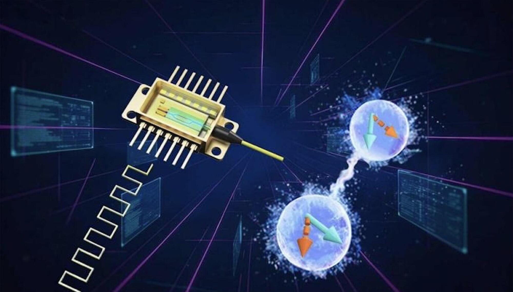
Quantum technologies are cutting-edge systems that can process, transfer, or store information leveraging quantum mechanical effects, particularly a phenomenon known as quantum entanglement. Entanglement entails a correlation between two or more distant particles, whereby measuring the state of one also defines the state of the others.

In recent years, quantum physicists and engineers have been trying to realize devices that operate leveraging the entanglement between individual particles of light (i.e., photons). The reliable operation of these devices relies on so-called entangled photon sources (EPSs), components that can generate entangled pairs of photons.

Researchers at University of Science and Technology of China, Jinan Institute of Quantum Technology, CAS Institute of Semiconductors and other institutes recently realized a new EPS integrated onto a single photonic chip, which can generate entangled photons via an electrically powered laser. Their study is published in Physical Review Letters.

Mercury’s BepiColombo Mio and Earth’s GEOTAIL show shared wave frequency properties across planetary magnetospheres

An international team from Kanazawa University (Japan), Tohoku University (Japan), LPP (France), and partners has demonstrated that chorus emissions, natural electromagnetic waves long studied in Earth’s magnetosphere, also occur in Mercury’s magnetosphere exhibiting similar chirping frequency changes.

Using the Plasma Wave Investigation instrument aboard BepiColombo’s Mercury orbiter Mio, six Mercury flybys between 2021 and 2025 detected plasma waves in the audible range. Comparison with decades of GEOTAIL data confirmed identical instantaneous frequency changes.

This provides the first reliable evidence of intense electron activity at Mercury, advancing understanding of auroral processes across the solar system.

Turning MRI into a quantitative microscope to detect white matter injury

Early diagnosis and noninvasive monitoring of neurological disorders require sensitivity to elusive cellular-level alterations that emerge much earlier than volumetric changes observable with millimeter-resolution medical imaging.

Morphological changes in axons—the tube-like projections of neurons that transmit electrical signals and constitute the bulk of the brain’s white matter—are a common hallmark of a wide range of neurological disorders, as well as normal development and aging.

A study from the University of Eastern Finland (UEF) and the New York University (NYU) Grossman School of Medicine establishes a direct analytical link between the axonal microgeometry and noninvasive, millimeter-scale diffusion MRI (dMRI) signals—diffusion MRI measures the diffusion of water molecules within biological tissues and is sensitive to tissue microstructure.

A genetic breakthrough links early-onset diabetes to brain disorders

Paediatric teams are now facing babies whose diabetes appears in the first weeks of life, then rapidly reveals deep problems in brain growth and function. A new genetic finding sheds light on how a single molecular fault can disrupt both blood sugar control and early brain development.

Neonatal diabetes is diagnosed in the first six months of life, often within days or weeks after birth. Unlike the more common type 1 diabetes, which usually shows up in children and teenagers, neonatal diabetes is almost always genetic.

Doctors typically notice poor feeding, weight loss, dehydration and extremely high blood sugar. In many cases, the root cause is a mutation that stops the pancreas from making enough insulin. That alone makes neonatal diabetes a medical emergency.

Recent discoveries on the acquisition of the highest levels of human performance

Scientists have long debated the origins of exceptional human achievements. This literature review summarizes recent evidence from multiple domains on the acquisition of world-class performance. We review published papers and synthesize developmental patterns of international top scientists, musicians, athletes, and chess players. The available evidence is highly consistent across domains: (i) Young exceptional performers and later adult world-class performers are largely two discrete populations over time. (ii) Early (e.g., youth) exceptional performance is associated with extensive discipline-specific practice, little or no multidisciplinary practice, and fast early progress. (iii) By contrast, adult world-class performance is associated with limited discipline-specific practice, increased multidisciplinary practice, and gradual early progress.

Human heart regrows muscle cells after heart attack, world-first study shows

This study provides the first direct evidence of cardiomyocyte mitosis in the adult human heart following myocardial infarction, challenging the long-standing paradigm that cardiac muscle cells are incapable of regeneration. Utilizing live human heart tissue models, researchers from the University of Sydney demonstrated that while fibrotic scarring occurs post-ischemia, the heart simultaneously initiates a natural regenerative program characterized by active cell division. The investigation further identified specific regulatory proteins that drive this mitotic process, offering a molecular blueprint for endogenous tissue repair. These findings suggest that the human heart possesses a latent regenerative capacity that could be therapeutically harnessed to prevent heart failure and reverse post-infarct tissue damage, representing a significant shift in regenerative cardiovascular medicine.


A world‑first University of Sydney study reveals that the human heart can regrow muscle cells after a heart attack, paving the way for breakthrough regenerative therapies to reverse heart failure.

Panoptic imaging of transparent mice reveals whole-body neuronal projections and skull–meninges connections

A nanobody-based immunolabeling method, vDISCO, boosts the signal of fluorescent proteins and allows imaging of subcellular details in intact transparent mice. It uncovers neuronal projections and skull–meninges connections in whole adult mice.

/* */