This website uses a security service to protect against malicious bots. This page is displayed while the website verifies you are not a bot.
Researchers developed a two-stage machine learning framework that detected diabetes and classified records as prediabetes, type 1, type 2, or type 3c diabetes using two public datasets. XGBoost showed strong internal performance, but inconsistent model rankings, non-standard variables, and the absence of external clinical validation limit immediate use.
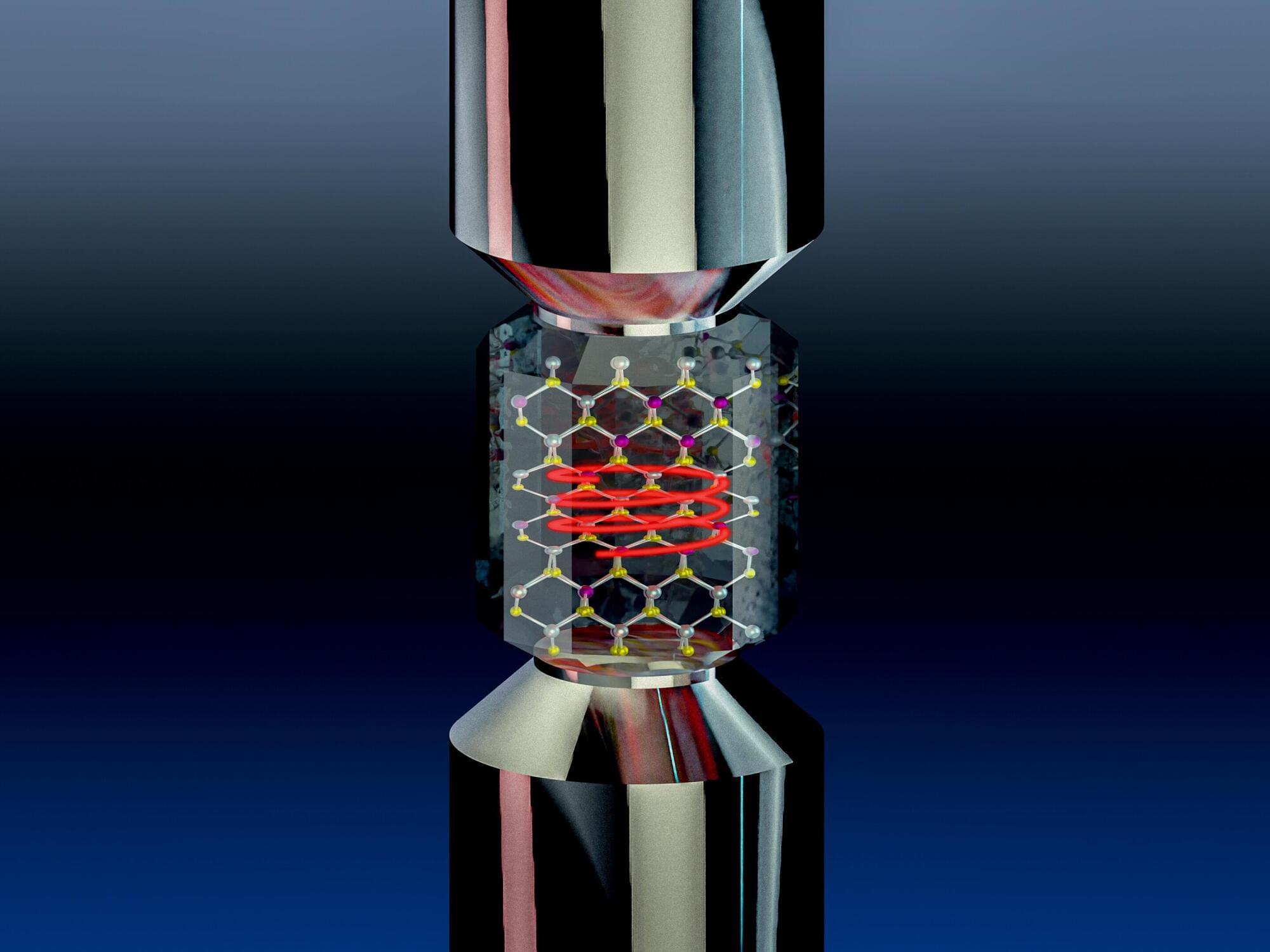
Mechanical strain is one of the most common tools used to tailor the properties of materials. In piezoelectric materials, stretching or compressing a crystal generates an electrical polarization. In piezomagnetic materials, it induces magnetization. Researchers at the Max Planck Institute for the Structure and Dynamics of Matter (MPSD) and the University of Oxford have now discovered that mechanical strain also induces chirality in non-chiral crystals, opening a new direction to control this property on demand and potentially imprint chiral electronic properties. This work has just been published in Nature.
Chirality is an important property of matter. It is defined as a property of objects that cannot be superimposed on their mirror images through any combination of rotations or translations, much like distinct left and right hands. In chiral crystals, the spatial arrangement of atoms gives rise to a specific handedness, with the crystal structure twisting along one propagation direction in a way similar to a screw.
As a consequence, propagation in one direction, for example, of an electrical current, may experience different resistance than propagation in the opposite direction. This effect also influences certain chemical reactions and biological processes, which select for one specific handedness. In this sense, the ability to control chirality on demand, turning a right-handed structure into a left-handed one, is highly desirable.
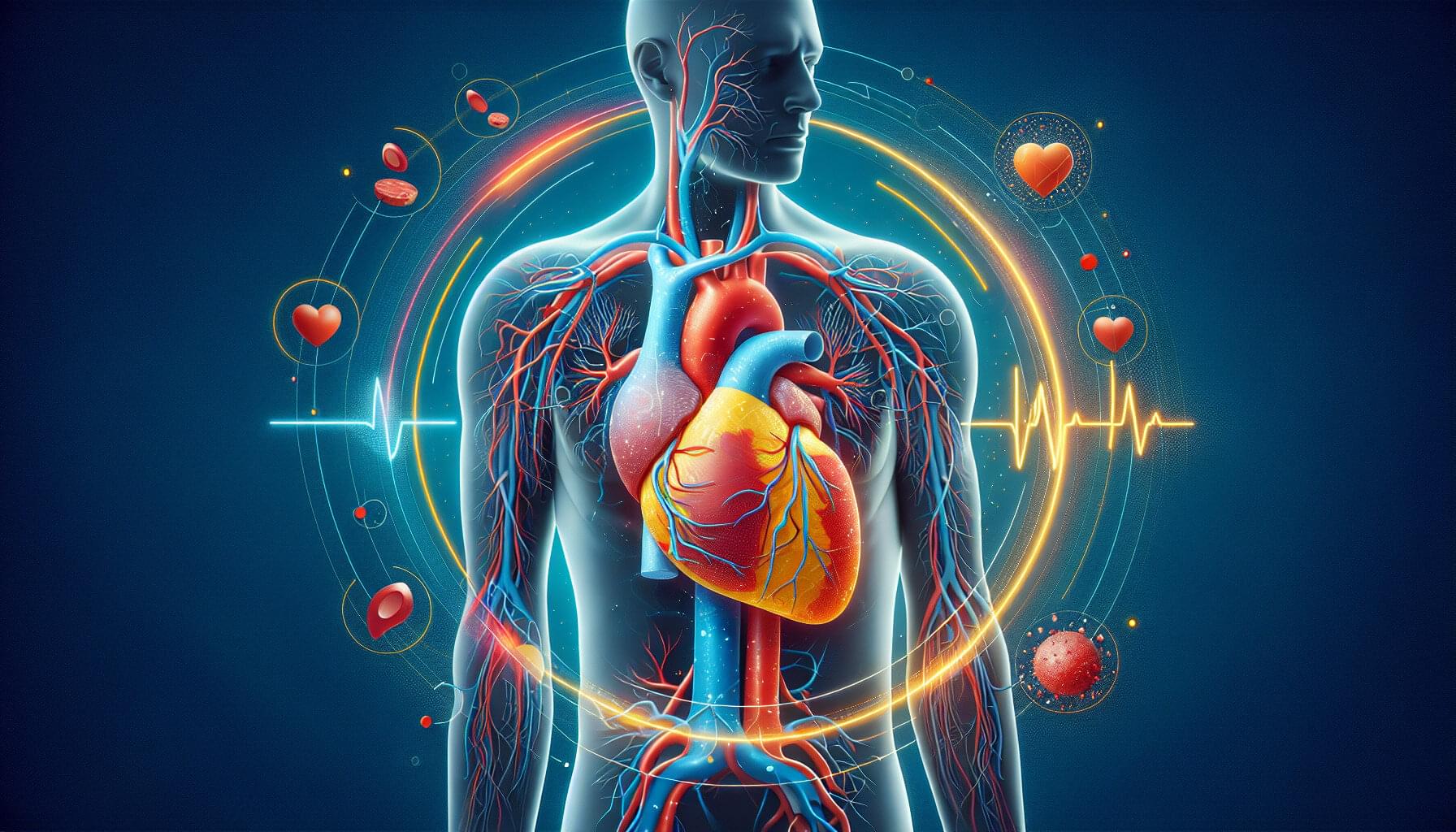
Specialized cells in the human body make biological trade-offs to perform certain jobs. To support the large, hardworking hearts that power other organs, heart cells have evolved to be extremely efficient and resilient. Because the adult human heart cannot repair itself the way skin does, scientists have studied ways to “reprogram” heart cells to regenerate after heart attacks. Heart cells have very stable identities, however, and are resistant to reprogramming.
Scientists at Sanford Burnham Prebys Medical Discovery Institute and collaborators at Johns Hopkins University School of Medicine published findings in Nature Communications that cover a new mechanism cells use to protect their identities and ward off reprogramming attempts. Continued study of the obstacles to cellular reprogramming may lead to treatments that can help the heart repair itself after injury.
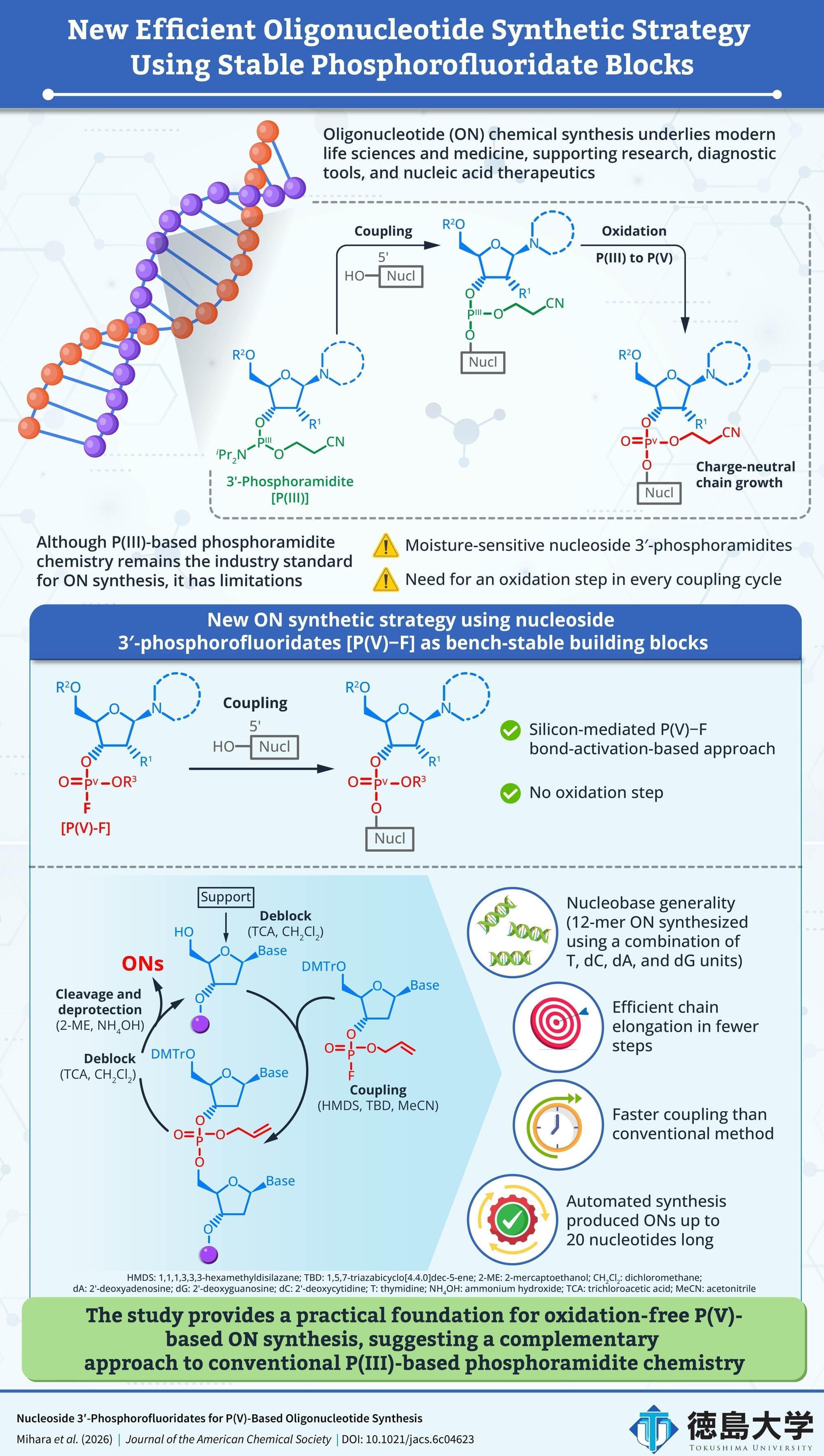
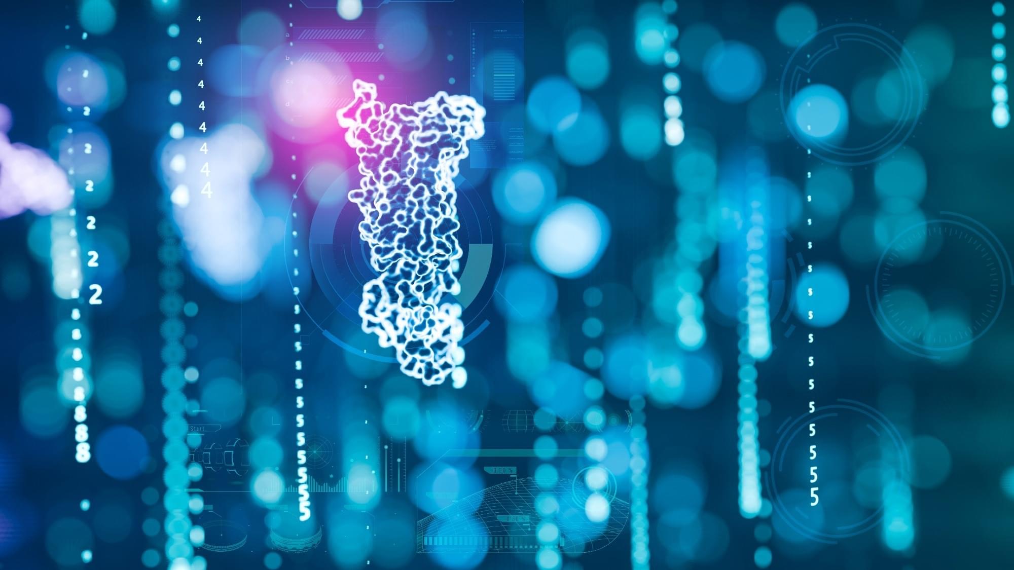
The chemical synthesis of oligonucleotides (ONs) is central to modern molecular biology, diagnostics and nucleic acid therapeutics. While demand for high-quality ONs is increasing, the conventional synthetic method has long-standing efficiency challenges. Widely adopted P(III)-phosphoramidite-based ON synthesis requires an oxidation step after every nucleotide coupling cycle and uses moisture-sensitive building blocks, adding complexity to the workflow and slowing the process.
Early studies of ON synthesis showed that pentavalent phosphorus [P(V)] chemistry could form linkages between nucleotides. However, practical limitations, including unstable intermediates, slow coupling, harsh deprotection or poor performance during chain elongation, prevented these methods from replacing P(III)-based phosphoramidite chemistry.
A recent study led by Associate Professor Noriko Saito-Tarashima of the Graduate School of Pharmaceutical Sciences at Tokushima University in Japan, along with Nana Mihara, a doctoral student from the same institution, investigated whether nucleoside 3′-phosphorofluoridates [P(V)–F] could be used as stable building blocks for ON synthesis without requiring a separate oxidation step.
As prediabetes advances toward diabetes, this delicate process can begin to break down. Misfolded and defective proteins accumulate inside cells, creating stress that can damage the pancreatic cells responsible for producing insulin.
Researchers from Sanford Burnham Prebys Medical Discovery Institute and the University of Michigan reported new details about this process on June 1, 2026, in the Proceedings of the National Academy of Sciences. Their findings reveal how insulin-producing cells coordinate protein folding and what happens when that system falls out of balance. The work suggests that strengthening the cellular machinery responsible for folding proteins could help protect these cells from damage.
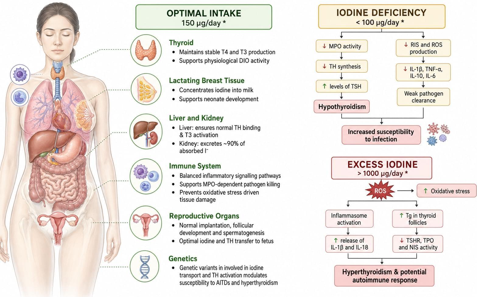
This narrative review finds that adequate iodine supports thyroid hormone signaling, leukocyte metabolism, antioxidant defenses, and antimicrobial activity in humans and domesticated mammals. Both deficiency and sustained excess may impair immunity or promote thyroid inflammation, but precise immune-specific intake thresholds remain uncertain.

Keep up to date on the latest data in advanced prostate cancer and gain expert insights on their implications for clinical care and personalized treatment.