A research team has developed the world’s first chip that can match the speed at which the human brain functions. The study, titled “A sub–10-millisecond neural dynamical system based on phase-change memristors,” was published in Science and was led by Professor Yang Yuchao of Peking University, together with researchers from the Shanghai Institute of Microsystem and Information Technology, Chinese Academy of Sciences.
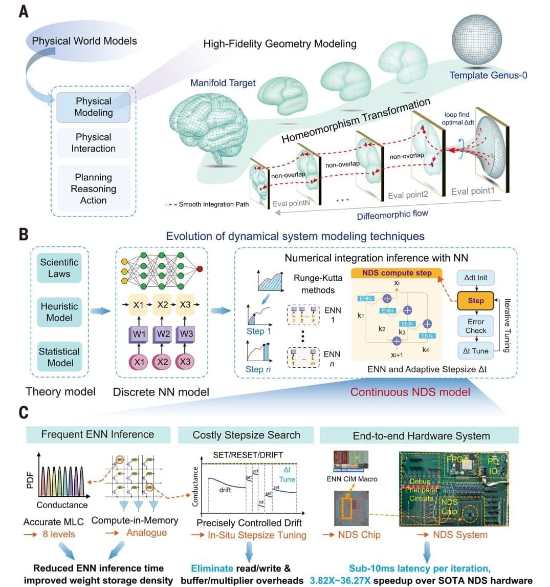
Neural dynamical systems combine neural networks with mathematical equations that describe how complex systems change over time. They are useful for physical modeling, medical imaging and three-dimensional brain reconstruction. However, these systems require repeated calculations, error checks and adjustments to the size of each calculation step. In conventional computers, data must also move frequently between memory and the processor, increasing processing time and energy use.
Fast and accurate brain modeling is important for technologies that must respond in real time, including brain–computer interfaces, surgical navigation and medical imaging. Existing hardware often requires too much time and power for these demanding calculations. By performing key operations directly in memory, the new chip reduces data movement and brings high-quality brain modeling closer to real-time use.