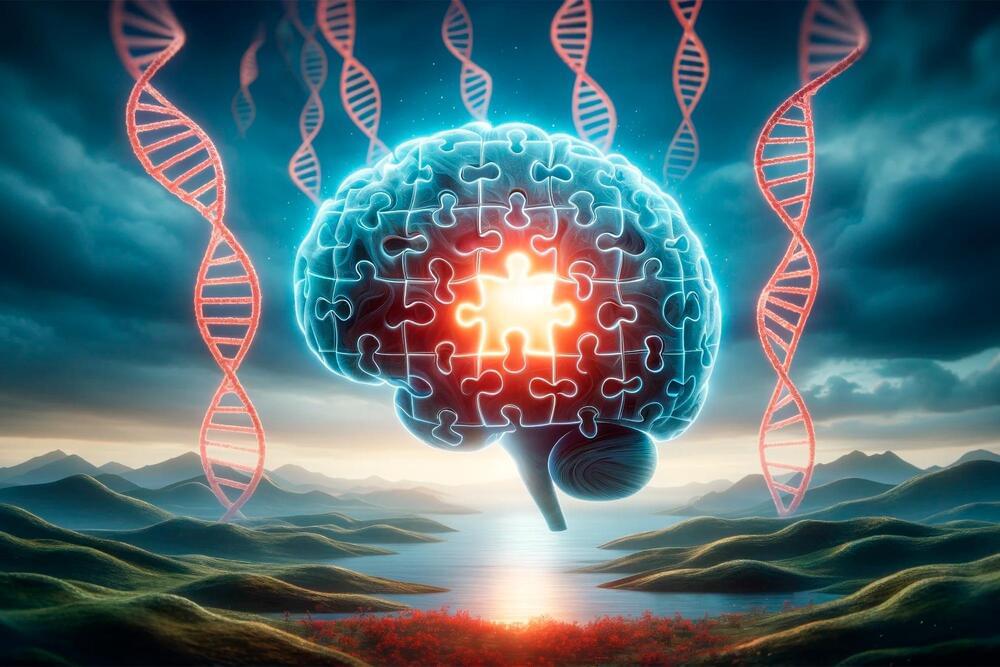
Breaking link between early, late stages of disease may prevent dementia.
Alzheimer’s disease has plagued one large Colombian family for generations, striking down half of its members in the prime of life. But one member of that family evaded what had seemed would be fate: Despite inheriting the genetic defect that caused her relatives to develop dementia in their 40s, she stayed cognitively healthy into her 70s.
For most adults, the majority of waking daily life is spent at work. That offers employers an opportunity to influence their employees’ physical, mental, social, and spiritual health.
To support the move to better health, the McKinsey Health Institute (MHI), along with other organizations such as the World Health Organization (WHO), are highlighting a more modern way to view health beyond illness and its absence.1 Adding years to life and life to years, McKinsey, March 29, 2022; A 2022 MHI survey on global health perspectives found that more than 40 percent of respondents who reported having a disease still perceived their health as good or very good, while more than 20 percent of those who reported no disease said they were in fair, poor, or very poor health. Embracing the concept of holistic health—an integrated view of an individual’s mental, physical, spiritual, and social functioning2 Previous work from MHI has defined each dimension of health in detail. For more details, see Adding years to life and life to years. Using this definition means that we emphasize “functioning.
This interview is an episode from @The-Well, our publication about ideas that inspire a life well-lived, created with the @JohnTempletonFoundation.
Watch Lisa Feldman Barrett’s next interview ► • The biggest myths about emotions, deb…
Our perception of reality is not an exact representation of the objective truth but rather a combination of sensory inputs and the brain’s interpretation of these signals. This interpretation is influenced by past experiences and is often predictive, with the brain creating categories of similar instances to anticipate future events.
The brain’s categorization process extends beyond physical characteristics to include abstract, functional features. This ability allows humans to create “social reality,” where we collectively assign functions or meanings to objects or concepts that don’t inherently possess them, such as the value of money or the concept of borders and citizenship.
The brain’s capacity for imagination, drawing from past experiences to create something entirely new, is a double-edged sword. While it allows for creativity and innovation, it can also lead to difficulties in staying present.
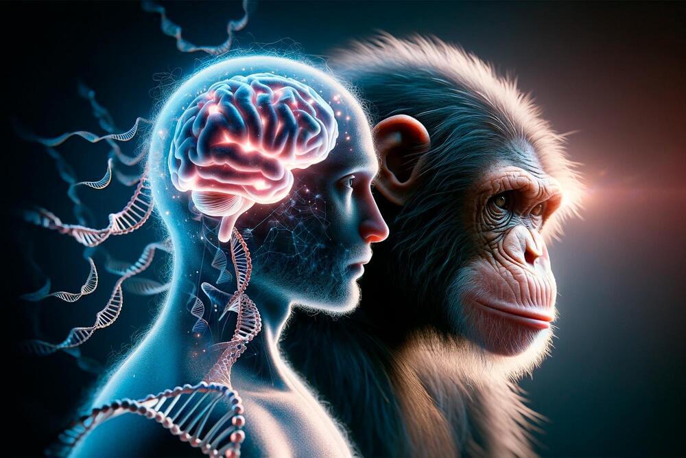
Primates are among the most intelligent creatures with distinct cognitive abilities. Their brains are relatively large in relation to their body stature and have a complex structure. However, how the brain has developed over the course of evolution and which genes are responsible for the high cognitive abilities is still largely unclear. The better our understanding of the role of genes in brain development, the more likely it will be that we will be able to develop treatments for serious brain diseases.
Researchers are approaching these questions by knocking out or activating individual genes and thus drawing conclusions about their role in brain development. To avoid animal experiments as far as possible, brain organoids are used as an alternative. These three-dimensional cell structures, which are only a few millimeters in size, reflect different stages of brain development and can be genetically modified. However, such modifications are usually very complex, lengthy and costly.
Researchers at the German Primate Center (DPZ)—Leibniz Institute for Primate Research in Göttingen have now succeeded in genetically manipulating brain organoids quickly and effectively. The procedure requires only a few days instead of the usual several months and can be used for organoids of different primate species. The brain organoids thus enable comparative studies of the function of genes at early stages of brain development in primates and help to better understand neurological diseases.
Thanks in part to Elon Musk, the field of brain-computer interfaces has captured both public and investor interest, with a cadre of companies now developing implantable devices.
The researchers found 139 genes that are common across the primate groups but highly divergent in their expression in human brains.
An international team led by researchers at the University of Toronto has uncovered over 100 genes that are common to primate brains but have undergone evolutionary divergence only in humans – and which could be a source of our unique cognitive ability.
The researchers, led by Associate Professor Jesse Gillis from the Donnelly Centre for Cellular and Biomolecular Research and the Department of Physiology at U of T’s Temerty Faculty of Medicine, found the genes are expressed differently in the brains of humans compared to four of our relatives – chimpanzees, gorillas, macaques, and marmosets.
This is not a work of art. It’s an image of microscopic blood flow in a rat’s brain, taken with one of many new tools that are yielding higher levels of detail in brain imaging.
Here are seven more glorious images from neuroscience research →
Facing an alcohol crisis, the US sees 12% of adult deaths linked to abuse. Excessive drinking risks permanent brain damage, Wernicke-Korsakoff Syndrome. Symptoms mimic drunkenness and can lead to irreversible psychosis. Prevention? Cut back or quit. Concerned? Seek medical advice for potential Vitamin B1 treatment.