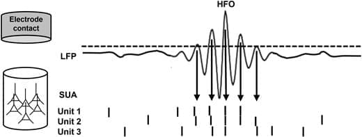
Toggle light / dark theme

Brain Dead Vs. Coma Vs. Vegetative State: What’s The Difference?

In light of Whitney Houston and Bobby Brown’s daughter, Bobbi Kristina being found unconscious, there have been many headlines that said she in “a coma” or a “vegetative state” or even “brain dead”. First things first, patients who suffer brain death are not in coma. And patients who are in coma may or may not progress to brain death.

The brain has a number of vast jobs to complete every second and is a very complex organ. The brain controls not only an individual’s thought process and voluntary movements, but it controls involuntary movements and other vital body functions. These functions include auditory, olfactory, visual and tactile senses, regulation of body temperature, blood pressure, respiration, and heart rate (although the heart can continue to beat without the brain in “autotonic response”). The brain also produces hormones to control individual organ function. A good example is the brain’s production of anti-diuretic hormone (ADH). This hormone is produced to concentrate the urine in the kidneys, thus protecting against life-threatening dehydration.

MUST READ: “Brain Dead” Child Showing Signs Of Life

Researchers discover how deadly brain cancer evades treatments

McMaster University researcher Sheila Singh and her team have discovered how glioblastoma, a lethal brain cancer, can evade treatments and kill.

The researchers found the cancer cells that survive the first round of radiotherapy or chemotherapy do so by mutating during the post-treatment minimal residual disease (MRD) or dormant state. The MRD profile of each patient was mapped using single cell sequencing to find a genetic signature that predicted how the cancer would recur in each individual.

Singh said that by mapping the MRD, researchers found that each patient had a different trajectory to their cancer recurring, potentially opening the door to future treatments tailored to each individual with glioblastoma. Singh’s team monitored five patients between 2018 and 2022.

We’ve just discovered a new part of the brain’s waste disposal system

In 2012, Nedergaard also helped to discover a network of thin tubes that collect waste fluid from brain cells, known as the glymphatic system. These tubes may drain into the outgoing cerebrospinal fluid, says Nedergaard.

The waste products of brain cells include proteins called beta-amyloid and tau that are thought to be involved in Alzheimer’s disease when they build up in excessive amounts.

In both mice and people, the SLYM also contains immune cells, so it may allow them to detect signs of infection present in the cerebrospinal fluid, says Nedergaard. “It is loaded with immune cells.”

A New Approach to Halting the Effects of Aging: Boosting Immune Cells Improves Brain Waste Clearance

Many neurodegenerative diseases, including Alzheimer’s.

Alzheimer’s disease is a disease that attacks the brain, causing a decline in mental ability that worsens over time. It is the most common form of dementia and accounts for 60 to 80 percent of dementia cases. There is no current cure for Alzheimer’s disease, but there are medications that can help ease the symptoms.

Jeff Lichtman (Harvard) Part 2: Neuromuscular Connectomics

The Connectome and Connectomics: Seeking Neural Circuit Motifs

Talk Overview: The human brain is extremely complex with much greater structural and functional diversity than other organs and this complexity is determined both by one’s experiences and one’s genes. In Part 1 of his talk, Lichtman explains how mapping the connections in the brain (the connectome) may lead to a better understanding of brain function. Together with his colleagues, Lichtman has developed tools to label individual cells in the nervous system with different colors producing beautiful and revealing maps of the neuronal connections.
Using transgenic mice with differently colored, fluorescently labeled proteins in each neuron (Brainbow mice), Lichtman and his colleagues were able to follow the formation and destruction of neuromuscular junctions during mouse development. This work is the focus of Part 2.
In Part 3, Lichtman asks whether some day it might be possible to map all of the neural connections in the brain. He describes the technical advances that have allowed him and his colleagues to begin this endeavor as well as the enormous challenges to deciphering the brain connectome.

Speaker Bio: Jeff Lichtman’s interest in how specific neuronal connections are made and maintained began while he was a MD-PhD student at Washington University in Saint Louis. Lichtman remained at Washington University for nearly 30 years. In 2004, he moved to Harvard University where he is Professor of Molecular and Cellular Biology and a member of the Center for Brain Science.
A major focus of Lichtman’s current research is to decode the map of all the neural connections in the brain. To this end, Lichtman and his colleagues have developed exciting new tools and techniques such as “Brainbow” mice and automated ultra thin tissue slicing machines.

Rafael Yuste: Can You See a Thought? Neuronal Ensembles as Emergent Units of Cortical Function

IBM Research hosts a fascinating seminar with Rafael Yuste, a world leader in optical methods for brain research. According to Professor Yuste, lifting neuroscience studies from looking at neurons one at a time to ensembles or functional units is key in our quest to understanding how brains work.

Rafael Yuste is a Professor of Biological Sciences and Neuroscience at Columbia University and Co-director of the Kavli Foundations Institute for Neural Circuitry. He recently helped launch the BRAIN Initiative, a large-scale scientific project to systematically record and manipulate the activity of complete neural circuits.

In this seminar, Rafael Yuste shows the results from his group’s two-photon holographic methods to selectively image and manipulate the activity of neuronal populations in 3D in vivo. These experiments – from imaging neuron activities, triggering of neuron ensemble activities, and even altering behavioral choices bi-directionally – are shedding light on the possibility of neuronal ensembles being functional building blocks of cortical circuits.

New Alzheimer’s Drug Approved by FDA, Promises to Slow Disease

U.S. health regulators gave early approval to a new Alzheimer’s drug from Eisai Co. and Biogen Inc., the most promising to date in a new class of medicines that may help slow cognitive decline caused by the disease.

The Food and Drug Administration granted conditional approval to the drug, called lecanemab, based on an early study finding it reduced levels of a sticky protein called amyloid from the brains of people with early-stage Alzheimer’s. The companies will sell it under the brand name Leqembi.

Scientists discover new anatomic structure in the brain that monitors and shields cells

Though the team largely explains the function of SLYM in mice, they do study its presence in the adult human brain as well.

The human brain is tremendously complex, and scientists are yet to unlock its full potential. Now, a discovery has identified a previously unknown component of brain anatomy that doubles up as a protective barrier for our grey matter and a platform from which immune cells can monitor the brain, according to a release.

Maiken Nedergaard, co-director of the Center for Translational Neuromedicine at the University of Rochester and the University of Copenhagen, and Kjeld Møllgård, M.D.


Nopparit/iStock.

The researchers named the layer SLYM, an abbreviation of Subarachnoidal LYmphatic-like Membrane. SLYM divides the space below the arachnoid layer and the subarachnoid space into two sections.