Enjoy the videos and music you love, upload original content, and share it all with friends, family, and the world on YouTube.
Category: neuroscience – Page 533
Mitochondria Dump DNA in The Brain, Potentially Cutting Years Off Our Lives
Scraps of DNA discarded by our neurons’ power units are being absorbed into our nuclear genome far more frequently than assumed, potentially putting our brains at greater risk of developing life-threatening conditions.
An investigation by a team of researchers led by Columbia University in the US has found individuals with higher numbers of nuclear mitochondrial insertions – or NUMTs (pronounced new-mites) – in their brain cells are more likely to die earlier than those with fewer DNA transfers.
Mitochondria serve as our cells’ batteries, churning out energy in a form of chemical currency that suits most of our body’s metabolic needs. Once a discrete microbial organism in its own right, these tiny powerhouses were co-opted by our unicellular ancestors billions of years in the past, genes and all.

Do Plastics Cause Autism? Here’s what the latest study really says
A study out recently has prompted much media attention about the role of plastics in developing autism.
In particular, the study focused on exposure to a component of hard plastics—bisphenol A, or BPA—in the womb and the risk of boys developing this neurodevelopmental disorder.
Importantly, the study doesn’t show plastics containing BPA cause autism.
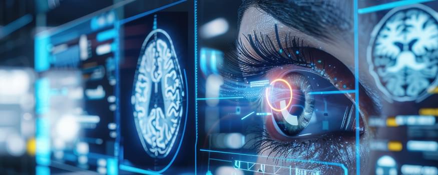
Diagnosing traumatic brain injury with a handheld device
The retina and optic nerve share most of the brain’s biochemical properties – this way, they provide a ‘window’ into the biochemistry of the brain.
To address this lack of technological means for the early detection of TBI, Pola Goldberg Oppenheimer, a Professor in Micro-Engineering and Bio-Nanotechnology at the University of Birmingham, UK, has developed a groundbreaking laser-based, eye-safe device (EyeD) technology. This technology can detect molecular changes that reflect brain damage by scanning the back of the eye with a handheld device.
Study Links Gene Variations to Brain Changes in Essential Tremor
Summary: Researchers identified how gene variations lead to brain changes associated with essential tremor, a common movement disorder affecting over 60 million people worldwide. The study used brain MRI scans and genetic data from over 33,000 adults to uncover genetic links to structural changes in the brain’s cortex and cerebellum.
These findings could lead to new drug targets by revealing how faulty protein disposal systems disrupt neural pathways, resulting in uncontrollable hand tremors. The research marks a significant step toward understanding and treating essential tremor more effectively.
Clinical Reasoning: A 50-Year-Old Man With Intracerebral Hemorrhage and Tortuous Retinal Arterioles
A 50-year-old man presented with headache. Examination showed left sided ataxic hemiparesis and elevated blood pressure. Brain imaging revealed an acute intracerebral hemorrhage in the right lentiform nucleus, deep and periventricular white matter hyperintensities, and predominantly deep cerebral microbleeds. Fundus examination showed important arteriolar tortuosity involving several blood vessels. In this young patient, we explain the diagnostic approach to intracerebral hemorrhage, the causes of cerebral small vessel disease, and the interpretation of biomolecular tests.
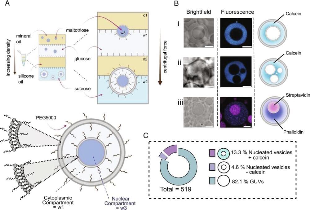
Nucleated synthetic cells with genetically driven intercompartment communication
The brain’s white matter comprises areas of the central nervous system made up of myelinated axons. Its name is derived from the pale appearance of the lipids that comprise myelin. Myelin is a segmented sheath that insulates axons, ensuring the conduction of neural signals. The loss of myelin is documented in a number of neurodegenerative pathologies, including Alzheimer’s and Parkinson’s disease, and perhaps most notably, multiple sclerosis. As people age, demyelination becomes more likely.
Researchers have long suspected a relationship between cardiorespiratory fitness and the integrity of the brain’s white matter as people age. However, a lack of specific evidence has led researchers at the National Institutes of Health to conduct a study examining the strength of this correlation, now published in the Proceedings of the National Academy of Sciences.
To establish a correlation between cardiovascular fitness and cerebral myelination, the researchers recruited a cohort of 125 participants from age 22 to 94 years old. The cardiovascular fitness of the participants was quantified as the maximum rate of oxygen consumption, popularly and succinctly known as VO2max. Myelin content was defined as the myelin water fraction, which the researchers estimated through an advanced multicomponent relaxometry MRI method.
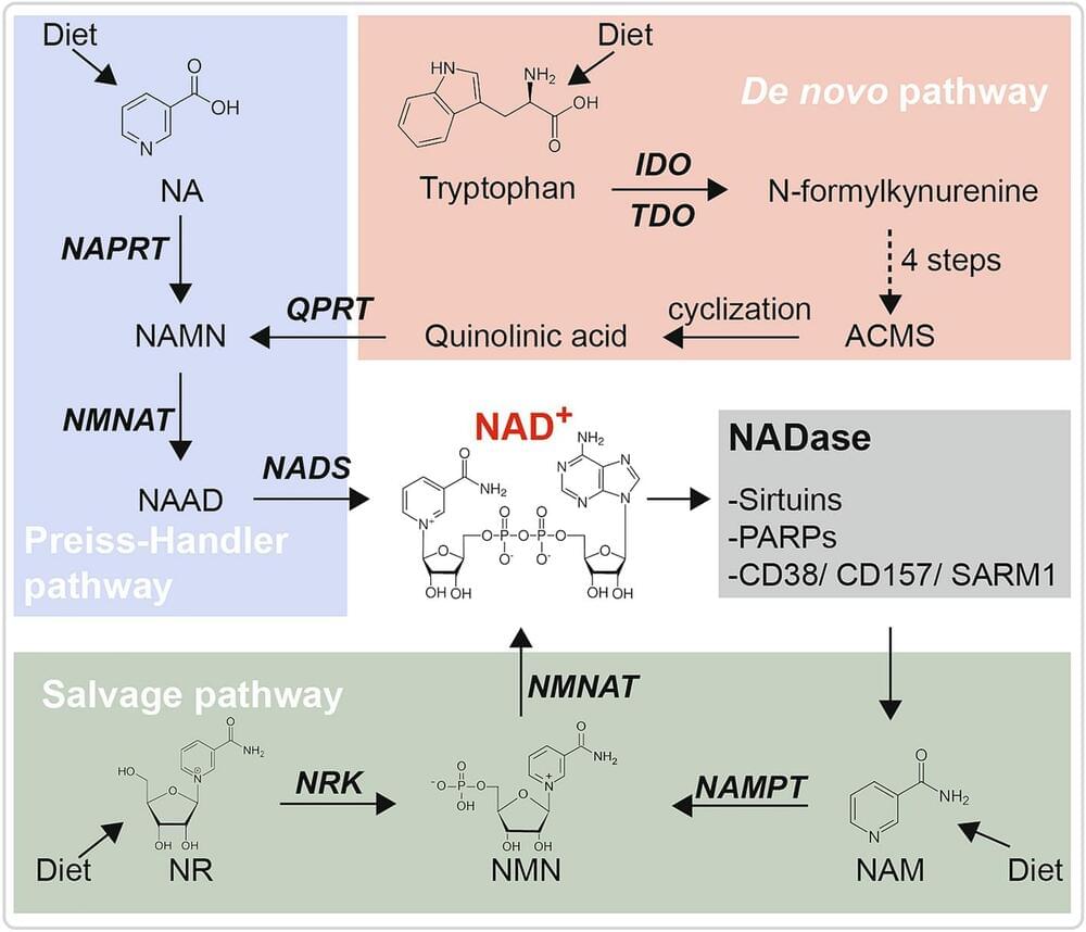
Targeting NAD Metabolism for the Therapy of Age-Related Neurodegenerative Diseases
Shan C, Gong YL, Zhuang QQ, Hou YF, Wang SM, Zhu Q, et al. Protective effects of β- nicotinamide adenine dinucleotide against motor deficits and dopaminergic neuronal damage in a mouse model of Parkinson’s disease. Prog Neuro Psychopharmacol Biol Psychiatry 2019, 94: 109670.

Humanity’s newest brain gains are most at risk from ageing
In the more than six million years since people and chimpanzees split from their common ancestor, human brains have rapidly amassed tissue that helps decision-making and self-control.
The large prefrontal cortex provides evolutionary and cognitive advantages over non-human primates — but there’s a cost.
Consciousness pre-dates life
Many scientists, operating with a materialist worldview, argue that consciousness emerges out of inanimate molecules. In contrast, Roger Penrose’s longtime collaborator, Stuart Hameroff, puts forward the controversial case that consciousness precedes life and that we have evidence for this from a recent NASA experiment.