‘Revolutionary breakthrough’ could allow for smartphone-free Google searches


Spaceflight makes certain human stem cells age faster, a new study has found, furthering scientists’ understanding of the potential effects of space exploration on the human body.
Stem cells are found throughout the body, and they can make more of themselves or turn into other specialized cells — including blood, brain or bone cells — for maintenance and repair.
“In space, stem cells decline in function,” said lead study author Catriona Jamieson, director of the Sanford Stem Cell Institute and professor of medicine at the University of California, San Diego School of Medicine. “They actually reduce their ability to renew themselves or regenerate, and that’s an important thing to be able to know for long-term space missions.”
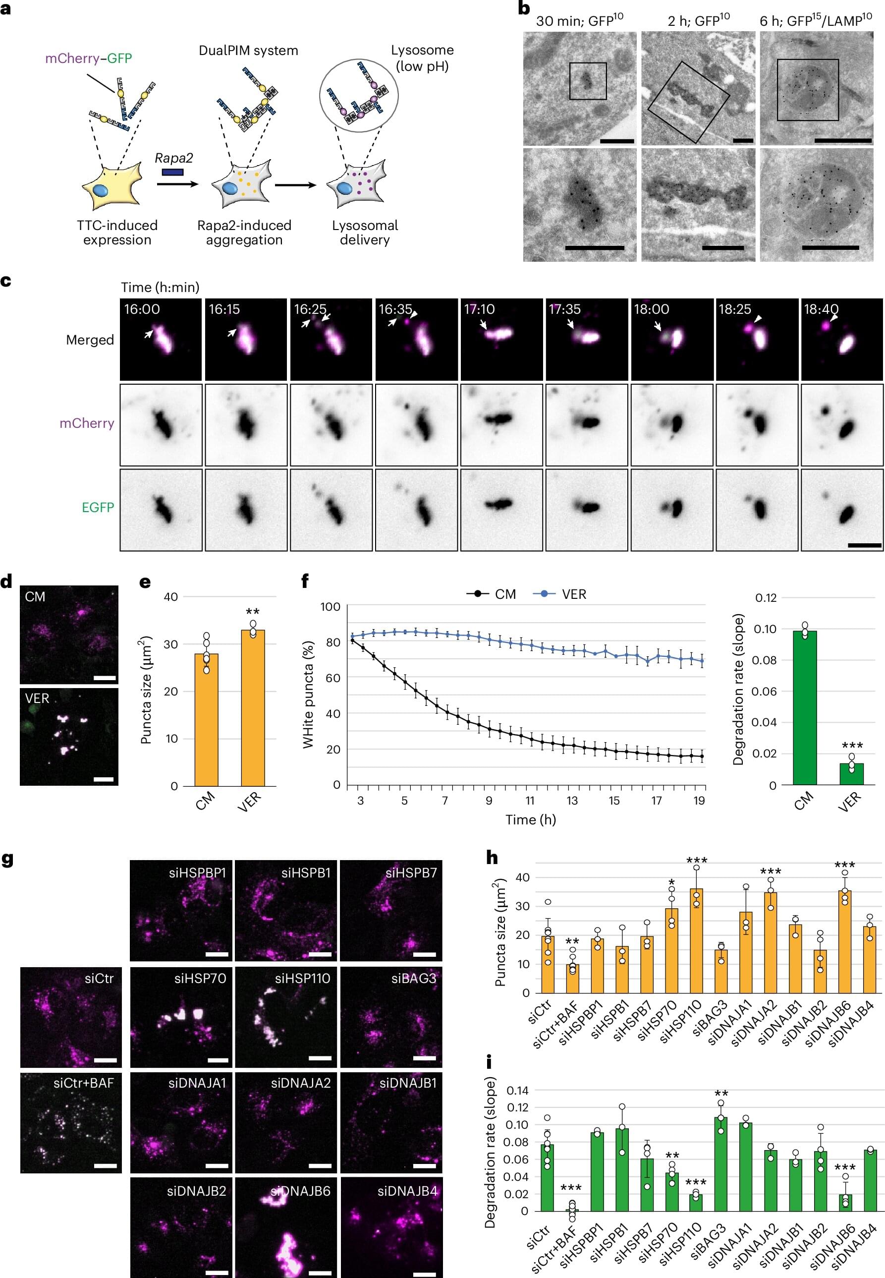
A new study from Aarhus University shows that our cells’ ability to clean out old protein clumps, known as aggregates, also includes a—up till now unknown—partnership with an engine that breaks down bigger pieces into smaller before “taking it to the trash.” An important find for future treatments of diseases like Alzheimer’s, Parkinson’s, ALS and Huntington’s, which are all characterized by the accumulation of protein in the brain.
Imagine you’re about to eat a big pizza. In order to not choke on it, you cut it up into slices and eat it bite by bite. And while you’re chomping down on your slices, cells inside your body are busy slicing the built-up protein clumps into pieces that are more manageable for the body’s trash system—otherwise it would clog up and malfunction.
Researchers from the department of Biomedicine at Aarhus University have just released a new study, which for the first time documents exactly how those clumps of unwanted protein get reduced to smaller pieces before being disposed of by the cells’ garbage disposal system—called autophagy. The work is published in the journal Nature Cell Biology.

To shed new light on the contribution of different brain regions and neural circuits to specific mental functions, neuroscientists and medical researchers rely on advanced imaging techniques and neural probes. These are electronic devices embedding electrodes, components that can measure the electrical impulses produced by neurons, which are known as spikes.
While existing probes have helped to map various networks of neurons and understand their functions, most of them have two-dimensional (2D) layouts. This is because they are made employing conventional semiconductor-based devices and fabrication strategies.
Researchers at Dartmouth College, University of Pittsburgh, Oklahoma State University and other institutes have developed a new approach that could enable the fabrication of soft three-dimensional (3D) neural probes on a large scale. Their proposed method, outlined in a paper published in Nature Electronics, entails the rolling of flat electronic devices into cylindrical 3D structures.

For those partnered up in a long-term relationship, studies have shown that different health characteristics can sometimes be shared across the couple – and that extends to psychiatric disorders, according to new research.
Based on an analysis of more than 6 million couples across Taiwan, Denmark, and Sweden, an international team of researchers found that people were significantly more likely to have the same psychiatric conditions as their partners than would be expected by chance.
Those conditions included schizophrenia, attention deficit hyperactivity disorder (ADHD), depression, autism, anxiety, bipolar disorder, obsessive-compulsive disorder (OCD), substance abuse, and anorexia nervosa.

Neuroscientists from 22 labs joined forces in an unprecedented international partnership to produce a landmark achievement: a neural map that shows activity across the entire brain during decision-making.
The data, gathered from 139 mice, encompass activity from more than 600,000 neurons in 279 areas of the brain — about 95% of the brain in a mouse. This map is the first to provide a complete picture of what happens across the brain as a decision is made.
“They have created the largest dataset anyone has ever imagined at this scale,” said Dr. Paul W. Glimcher, chair of the department of neuroscience and physiology and director of the Neuroscience Institute at New York University’s Grossman School of Medicine, of the researchers.

UC Berkeley researchers mapped the brain circuits that control growth hormone during sleep, uncovering a feedback system where sleep fuels hormone release, and the hormone regulates wakefulness. The discovery helps explain links between poor sleep, obesity, diabetes, and cognitive decline, while opening new paths for treating sleep and metabolic disorders.

In fact, they age “ten times faster in space than on the ground,” said Dr. Catriona Jamieson, the director of the Sanford Stem Cell Institute at the University of California, San Diego, a lead author of the study.
Stem cells are special cells that can develop into various kinds of tissue. Stem cell aging is potentially worrisome because it diminishes the body’s natural ability to repair its tissues and organs, potentially leading to chronic, age-related conditions like cancer, neurodegenerative diseases and heart problems.
A paper in Molecular Biology and Evolution finds that the relatively high rate of autism-spectrum disorders in humans is likely due to how humans evolved in the past. The paper is titled “A general principle of neuronal evolution reveals a human accelerated neuron type potentially underlying the high prevalence of autism in humans.”
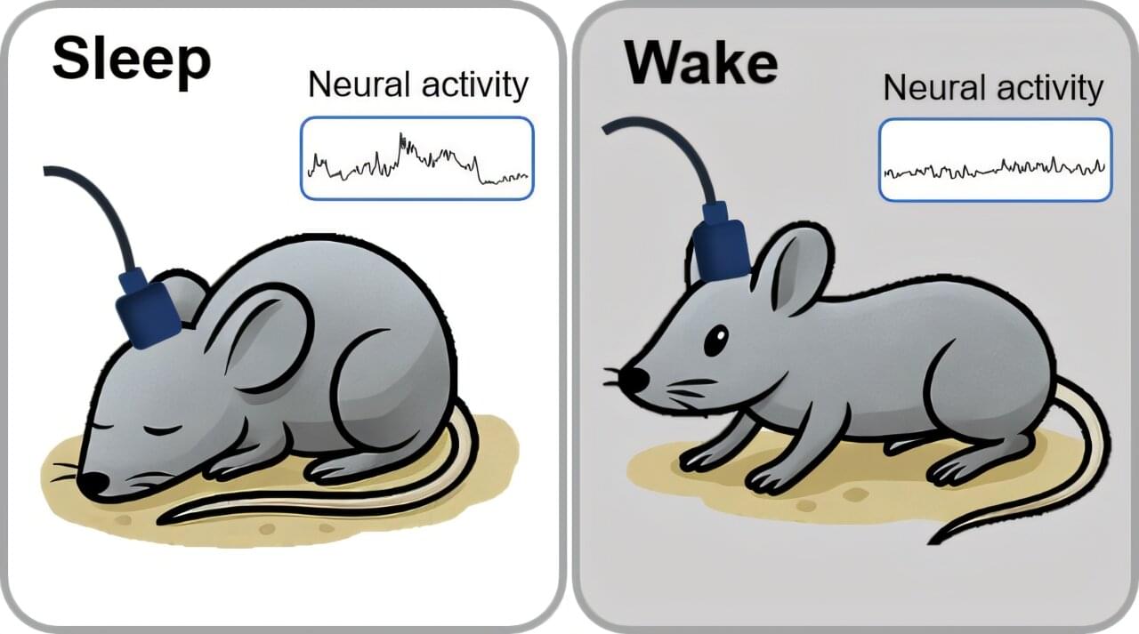
As every bodybuilder knows, a deep, restful sleep boosts levels of growth hormone to build strong muscle and bone and burn fat. And as every teenager should know, they won’t reach their full height potential without adequate growth hormone from a full night’s sleep.
But why lack of sleep —in particular the early, deep phase called non-REM sleep—lowers levels of growth hormone has been a mystery.
In a study published in Cell, researchers from University of California, Berkeley, dissect the brain circuits that control growth hormone release during sleep and report a novel feedback mechanism in the brain that keeps growth hormone levels finely balanced.