Western theories of consciousness tend to assume consciousness is about information.


Edward Chang is a neurosurgeon, scientist, and a pioneering leader in functional neurosurgery and brain-computer interface technology, whose work spans the operating room, the research lab, and the engineering bench to restore speech and movement for patients who have lost these capabilities. In this episode, Edward explains the evolution of modern neurosurgery and its dramatic reduction in collateral damage, the experience of awake brain surgery, real-time mapping to protect critical functions, and the split-second decisions surgeons make. He also discusses breakthroughs in brain-computer interfaces and functional electrical stimulation systems, strategies for improving outcomes in glioblastoma, and his vision for slimmer, safer implants that could turn devastating conditions like ALS, spinal cord injury, and aggressive brain tumors into more manageable chronic illnesses.
View show notes here: https://bit.ly/46uJXlh.
Become a member to receive exclusive content: https://peterattiamd.com/subscribe/
Sign up to receive Peter’s email newsletter: https://peterattiamd.com/newsletter/
We discuss:
0:00:00 — Intro.
0:01:17 — The evolution of neurosurgery and the shift toward minimally invasive techniques.
0:10:58 — Glioblastomas: biology, current treatments, and emerging strategies to overcome its challenges.
0:17:39 — How brain mapping has advanced from preserving function during surgery to revealing how neurons encode language and cognition.
0:24:22 — How awake brain surgery is performed.
0:29:02 — How brain redundancy and plasticity allow some regions to be safely resected, the role of the corpus callosum in epilepsy surgery, and the clinical and philosophical implications of disconnecting the hemispheres.
0:43:46 — How neural engineering may restore lost functions in neurodegenerative disease, how thought mapping varies across individuals, and how sensory decline contributes to cognitive aging.
0:54:40 — Brain–computer interfaces explained: EEG vs. ECoG vs. single-cell electrodes and their trade-offs.
1:09:02 — Edward’s clinical trial using ECoG to restore speech to a stroke patient.
1:20:41 — How a stroke patient regained speech through brain–computer interfaces: training, AI decoding, and the path to scalable technology.
1:41:10 — Using brain-computer interfaces to restore breathing, movement, and broader function in ALS patients.
1:47:56 — The 2030 outlook for brain–computer interfaces.
1:52:35 — The potential of stem cell and cell-based therapies for regenerating lost brain function.
1:57:54 — Edward’s vision for how neurosurgery and treatments for glioblastoma, Parkinson’s disease, and Alzheimer’s disease may evolve by 2040
2:00:43 — The rare but dangerous risk of vertebral artery dissections from chiropractic neck adjustments and high-velocity movements.
2:02:31 — How Harvey Cushing might view modern neurosurgery, and how the field has shifted from damage avoidance to unlocking the brain’s functions.
——-
About:
The Peter Attia Drive is a deep-dive podcast focusing on maximizing longevity, and all that goes into that from physical to cognitive to emotional health. With over 90 million episodes downloaded, it features topics including exercise, nutritional biochemistry, cardiovascular disease, Alzheimer’s disease, cancer, mental health, and much more.
Peter Attia is the founder of Early Medical, a medical practice that applies the principles of Medicine 3.0 to patients with the goal of lengthening their lifespan and simultaneously improving their healthspan.

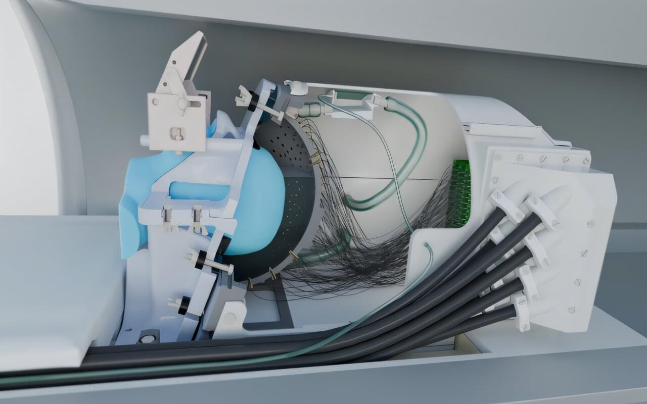
An ultrasound device that can precisely stimulate areas deep in the brain without surgery has been developed by researchers from UCL and the University of Oxford, opening up new possibilities for neurological research and treatment of disorders such as Parkinson’s disease.
A paper describing this work appears in Nature Communications.
Scientists have long been looking for a way to modulate brain function, which could improve our understanding of how the brain works and help to treat neurological diseases, using non-invasive methods that don’t involve surgery. One technology that could help is transcranial ultrasound stimulation (TUS), which was recently discovered to be able to modulate the activity of neurons (the brain’s key communication cells) by delivering gentle mechanical pulses that influence how these cells send signals.
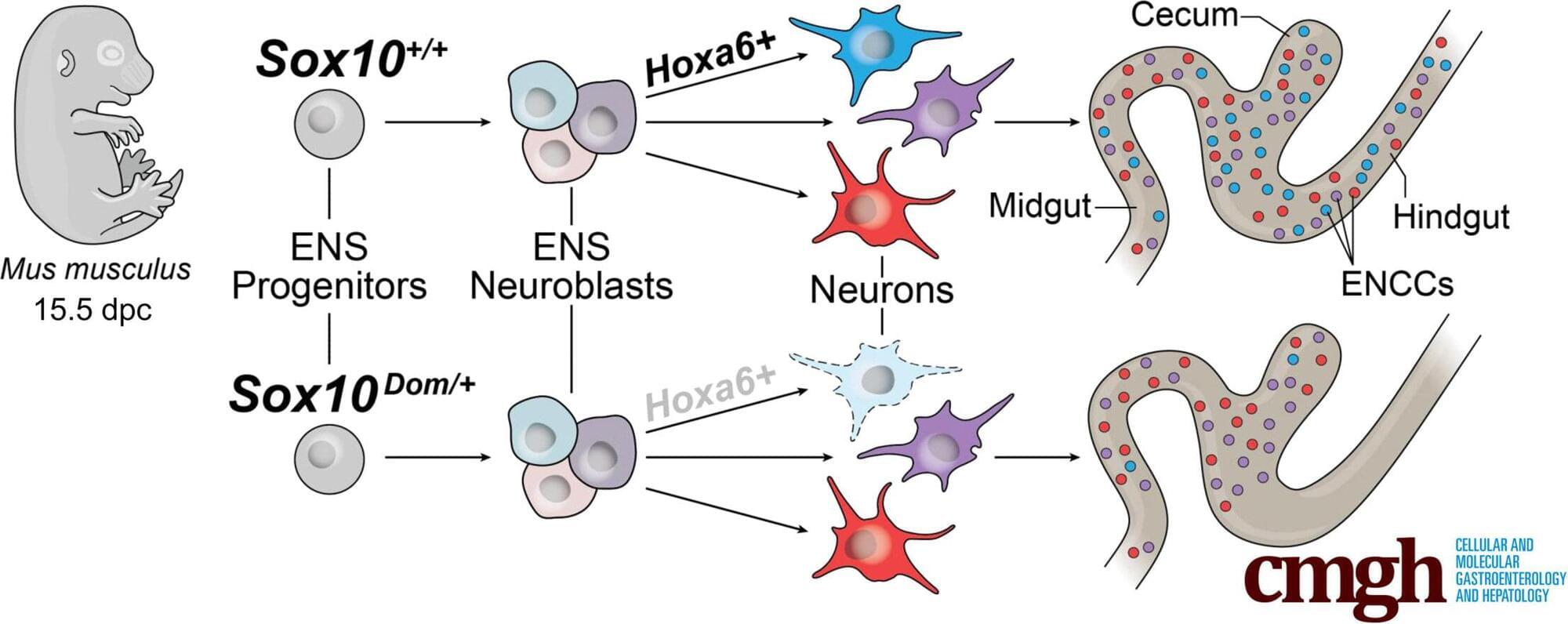
Vanderbilt researchers, including those from the Vanderbilt Brain Institute, have made significant strides in understanding how the enteric nervous system—sometimes called the “brain” of the gut—forms and functions.
In a study published in Cellular and Molecular Gastroenterology and Hepatology, the lab of principal investigator, Michelle Southard-Smith, sheds light on how the SOX10 protein contributes to the development of gut cells that play a role in gastrointestinal motility, or how food moves through the digestive system.
The paper is titled “Single Cell Profiling in the Sox10Dom Hirschsprung Mouse Implicates Hox genes in Enteric Neuron Trajectory Allocation.”
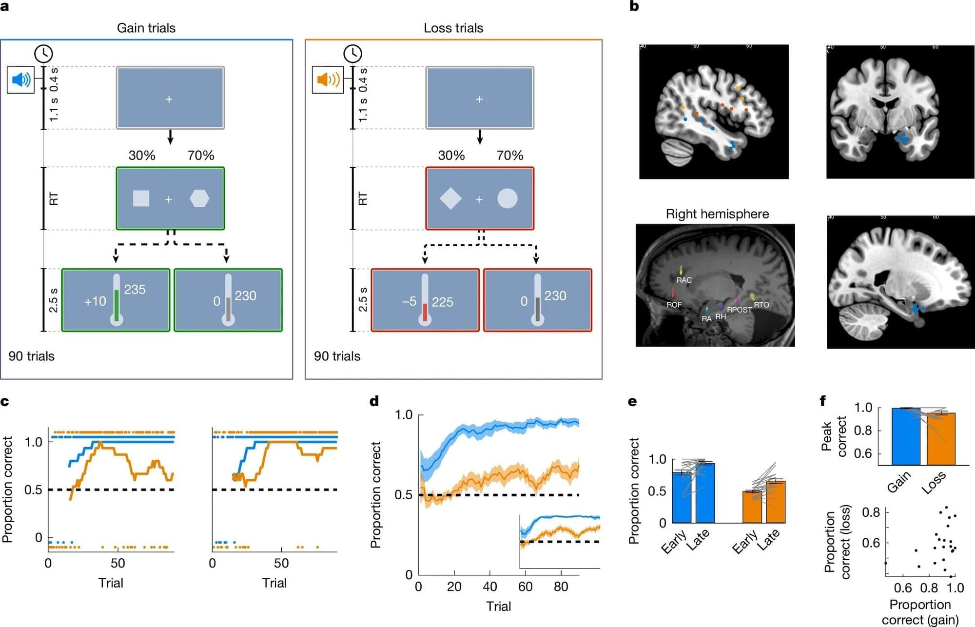
Researchers at the Weizmann Institute of Science traced a neural mechanism that explains why humans explore more aggressively when avoiding losses than when pursuing gains. Their work reveals how neuronal firing and noise in the amygdala shape exploratory decision-making.
Human survival has its origins in a delicate balance of exploration versus exploitation. There is safety in exploiting what is known, the local hunting grounds, the favorite foraging location, the go-to deli with the familiar menu. Exploitation also involves the risk of over-reliance on the familiar to the point of becoming too dependent upon it, either through depletion or a change in the stability of local resources.
Exploring the world in the hope of discovering better options has its own set of risks and rewards. There is the chance of finding plentiful hunting grounds, alternative foraging resources, or a new deli that offers a fresh take on old favorites. And there is the risk that new hunting grounds will be scarce, the newly foraged berries poisonous, or that the meal time will be ruined by a deli that disappoints.