Toggle light / dark theme

Radon, a naturally occurring radioactive gas produced when metals like uranium or radium break down in rocks and soil, is a known cause of lung cancer. Now new research has found exposure to high levels of this indoor air pollutant is associated with an increased risk of another condition in middle age to older female participants with ischemic stroke. The study is published in the January 3, 2024, online issue of Neurology, the medical journal of the American Academy of Neurology. Ischemic stroke is caused by a blockage of blood flow to the brain and is the most common type of stroke.

The condition, called clonal hematopoiesis of indeterminate potential (CHIP), develops when some hematopoietic stem cells, the building blocks for all blood cells, undergo genetic mutations as a person ages. Cells with such mutations replicate more quickly than cells without them. Previous research has shown people with CHIP may have a higher risk of blood cancers like leukemia and cardiovascular disease including stroke.

The study involved 10,799 female participants with an average age of 67. Approximately half of participants had a stroke or blood clots.

Nearly 2 million Americans suffer from type 1 diabetes — a condition that causes drastic spikes or drops in sugar levels and, in turn, dizziness, nausea, and fatigue. It’s a condition that must constantly be monitored, something that a lot of diabetics find mentally exhausting.

One diabetic, Naomi, told the BBC that she couldn’t handle “the physical or mental challenges of diabetes anymore,” and struggled to monitor her blood sugar levels multiple times a day. Naomi’s struggle isn’t unique — it’s called diabetes burnout.

There’s no cure for type 1 diabetes. However, researchers at the University of Arizona have adapted a cancer immunotherapy technique that has produced promising results in treating diabetes (in mice). The researchers engineered immune cells to fight off rogue T cells (immune cells that go haywire and attack the body) that can damage the pancreas, causing type 1 diabetes.

Several techniques currently are used to determine the pace of aging in animals and, to a lesser degree, in humans. However, the techniques used in humans lack accuracy, don’t assess aging in specific organs, are not widely available, and are expensive.

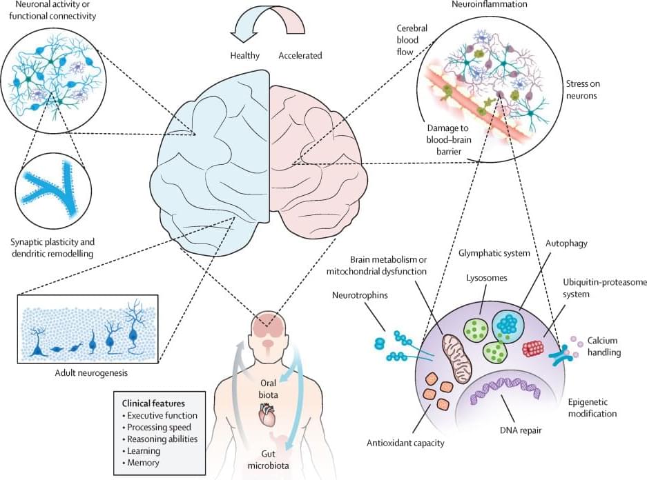
A multi-institutional research team measured the levels of nearly 5,000 human proteins in 5,676 people of all ages who were followed for as long as 15 years in five prospective longitudinal cohorts. Each measured protein was associated with specific organs, based on previous studies: adipose tissue, artery, brain, heart, immune tissue, intestine, kidney, liver, lung, muscle, or pancreas. Combinations of proteins indicated the pace of aging in each organ. Accelerated aging of one organ was found in nearly 20% of people, and accelerated aging of multiple organs was noted in ≈2%. Accelerated aging in a specific organ correlated with risk for developing disease in that organ. For example, people with accelerated heart aging (vs. those without it) had 250% higher risk for developing heart failure, and people with accelerated brain and vascular aging had nearly 60% higher risk for developing Alzheimer disease.

Various tools — from sequencing a person’s genome to measuring gene expression (e.g., the “methylome”) — are becoming available to predict a person’s risk for developing particular diseases. Will these predictions lead to interventions that lower risk? The jury is still out on that question.

In a small group of veterans diagnosed with mild traumatic brain injury, treatment with a psychedelic drug, ibogaine was associated with improvements in daily function and mental health symptoms, a new study out of Stanford found.

“This could be one of the first treatments for traumatic brain injury,” said Dr. Nolan Williams, associate professor of psychiatry at Stanford, and principal study investigator. “I think it’s a moment of hope for veterans and folks with permanent neurological injury.”

The Federal Drug Administration classifies ibogaine as a Schedule I drug, citing “high abuse potential” and “no accepted medical use.” To receive the one-time dose, 30 Special Ops veterans traveled to a treatment site in Mexico where ibogaine use is unregulated.

How deeply someone can be hypnotized — known as hypnotizability — appears to be a stable trait that changes little throughout adulthood, much like personality and IQ. But now, for the first time, Stanford Medicine researchers have demonstrated a way to temporarily heighten hypnotizablity — potentially allowing more people to access the benefits of hypnosis-based therapy.

In the new study, published Jan. 4 in Nature Mental Health, the researchers found that less than two minutes of electrical stimulation targeting a precise area of the brain could boost participants’ hypnotizability for about one hour.

“We know hypnosis is an effective treatment for many different symptoms and disorders, in particular pain,” said Afik Faerman, PhD, a postdoctoral scholar in psychiatry and lead author of the study. “But we also know that not everyone benefits equally from hypnosis.”

Over the past decades, scientists have made substantial progress unveiling the underlying mechanisms behind many psychiatric disorders. Every year, new genetic mutations or protein dysregulations are identified as potential culprits for the symptoms and sometimes even the root causes of complex neurological diseases, including autism spectrum disorder (ASD), schizophrenia, and Alzheimer’s.

Despite these efforts, the precise roles of several proteins involved in remain obscure. Such is the case for indoleamine 2,3-dioxygenase 2 (IDO2), an enzyme expressed in the brain and metabolized by the tryptophan–kynurenine pathway (TKP).

Changes in the metabolites of this pathway have already been linked to many , and genetically modified mice have been invaluable tools in such studies. However, the detailed functions of IDO2 in the brain are not known.

Human decision-making has been the focus of a wide range of research studies. Collectively, these research efforts could help to understand better how people make different types of everyday choices while also shedding light on the neural processes underpinning these choices.

Findings suggest that while making instantaneous decisions, or in other words, choices that need to be made quickly based on the information available at a given moment, humans greatly rely on contextual information. This contextual information can also guide so-called sequential decisions, which entails making a choice after observing the sequential unfolding of a process.

Researchers at the University of Oxford, the National Research Council in Rome, University College London (UCL), and the Max Planck Institute for Human Development recently carried out a study exploring the impact of context on goal-directed decision-making. Their findings, published in Neuron, suggest that goal-seeking ‘compresses’ spatial maps in the hippocampus and orbitofrontal cortices in the brain.

Whether in the brain or in the muscles, synapses are present wherever nerve cells exist. Synapses, the connections between neurons, are fundamental to the process of excitation transmission, which is essentially communication between neurons. As in any communication process, there is a sender and a receiver: Nerve cell processes called axons generate and transmit electrical signals thereby acting as signal senders.

Synapses are points of contact between axonal nerve terminals (the pre-synapse) and post-synaptic neurons. At these synapses, the electrical impulse is converted into chemical messengers that are received and sensed by the post-synapses of the neighboring neuron. The messengers are released from special membrane sacs called synaptic vesicles.

As well as transmitting information, synapses can also store information. While the structure and function of synapses are comparably well understood, little is known about how they are formed.