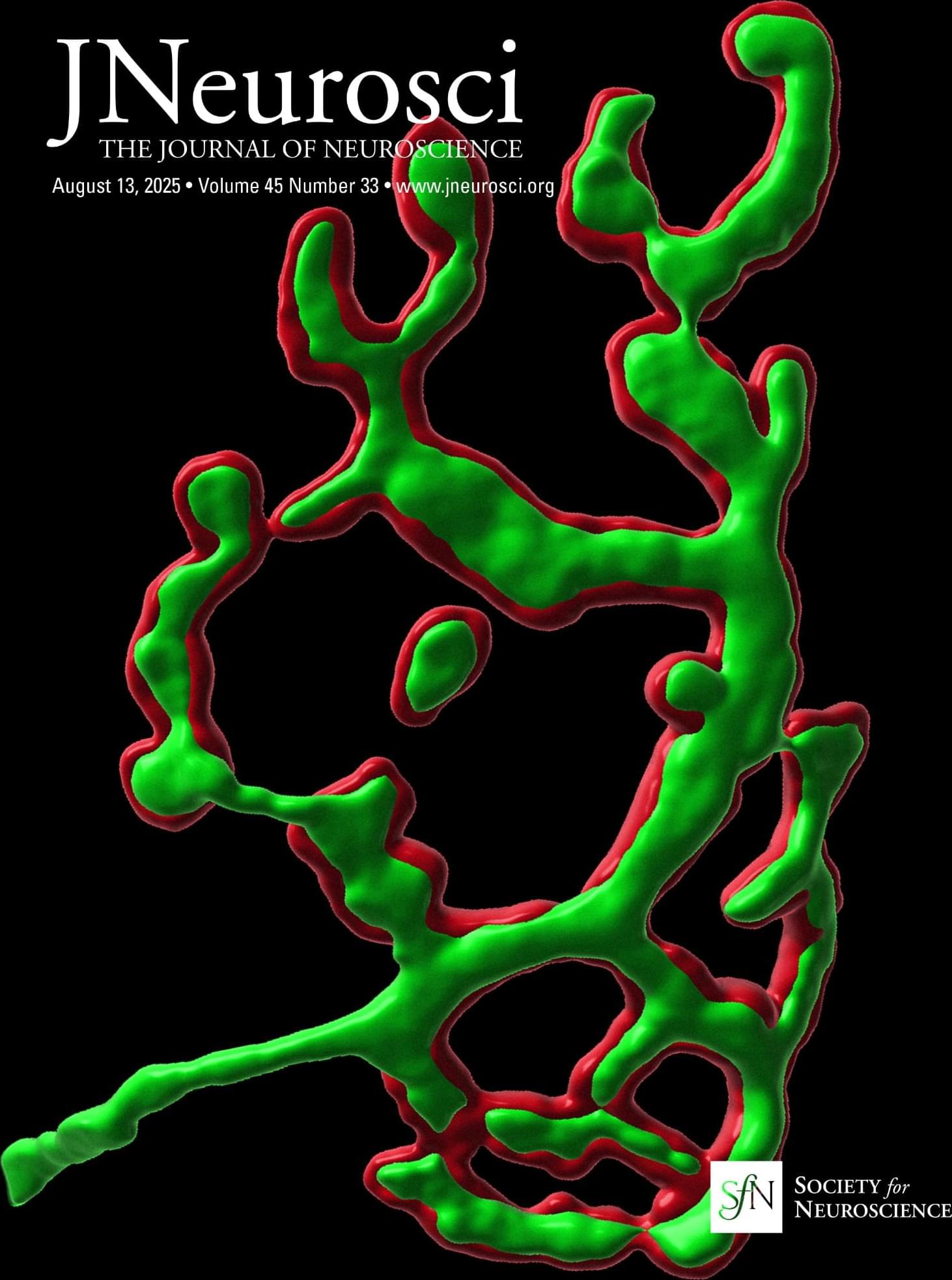
Toggle light / dark theme

A Novel 14mer Peptide Inhibits Autophagic Flux via Selective Activation of the mTORC1 Signalling Pathway: Implications for Alzheimer’s Disease

💡 LastestPaper.

📚 🔗 https://brnw.ch/21wPhFj.

🧑🏻‍🔬 By Ms. Cloe García Porta, Dr. Kashif Mahfooz, Mr. Joanna Komorowska, Dr. Sara Garcia-Rates and Dr. Susan Greenfield.


During development, a 14mer peptide, T14, modulates cell growth via the α-7 nicotinic acetylcholine receptor (α7 nAChR). However, this process could become excitotoxic in the context of the adult brain, leading to pathologies such as Alzheimer’s disease (AD). Recent work shows that T14 acts selectively via the mammalian target of rapamycin complex 1 (mTORC1). This pathway is essential for normal development but is overactive in AD. The triggering of mTORC1 has also been associated with the suppression of autophagy, commonly observed in ageing and neurodegeneration. We therefore investigated the relationship between T14 and autophagic flux in tissue cultures, mouse brain slices, and human Alzheimer’s disease hippocampus.

Brain mapping technique reveals insights into brain’s higher functions

A new way of mapping activity and connections between different regions of the brain has revealed fresh insights into how higher order functions like language, thought and attention, are organized.

Traditional models of activity represent interactions in pairs between two different brain regions. This is because modeling methods have not developed sufficiently to describe more between multiple regions.

A new approach, developed by researchers at the University of Birmingham is capable of taking signals measured through neuroimaging, and creating accurate models from these to show how different are contributing to specific functions and behaviors. The results are published in Nature Communications.

Network-based analyses uncover how neuroinflammation-causing microglia in Alzheimer’s disease form

Cleveland Clinic Genome Center researchers have unraveled how immune cells called microglia can transform and drive harmful processes like neuroinflammation in Alzheimer’s disease. The study, published in the journal Alzheimer’s & Dementia, also integrates drug databases with real-world patient data to identify FDA-approved drugs that may be repurposed to target disease-associated microglia in Alzheimer’s disease without affecting the healthy type.

The researchers, led by study corresponding author Feixiong Cheng, Ph.D., hope their unique approach of integrating genetic, chemical and human health data to identify and corresponding drugs will inspire other scientists to take similar approaches in their own research.

Microglia are specialized that patrol our brains, seeking and responding to tissue damage and external threats like bacteria and viruses. Different types of microglial cells use different methods to keep the brain safe. Some may cause neuroinflammation—inflammation in the brain—to fight invaders or kickstart the repair process in damaged cells. Others may work to “eat” dangerous substances in the brain, and clean up damage and debris. However, during Alzheimer’s disease, new types of microglia can form that promote .

Chaperone-mediated autophagy as a modulator of aging and longevity

Chaperone-mediated autophagy (CMA) is the lysosomal degradation of individually selected proteins, independent of vesicle fusion. CMA is a central part of the proteostasis network in vertebrate cells. However, CMA is also a negative regulator of anabolism, and it degrades enzymes required for glycolysis, de novo lipogenesis, and translation at the cytoplasmic ribosome. Recently, CMA has gained attention as a possible modulator of rodent aging. Two mechanistic models have been proposed to explain the relationship between CMA and aging in mice. Both of these models are backed by experimental data, and they are not mutually exclusionary. Model 1, the “Longevity Model,” states that lifespan-extending interventions that decrease signaling through the INS/IGF1 signaling axis also increase CMA, which degrades (and thereby reduces the abundance of) several proteins that negatively regulate vertebrate lifespan, such as MYC, NLRP3, ACLY, and ACSS2. Therefore, enhanced CMA, in early and midlife, is hypothesized to slow the aging process. Model 2, the “Aging Model,” states that changes in lysosomal membrane dynamics with age lead to age-related losses in the essential CMA component LAMP2A, which in turn reduces CMA, contributes to age-related proteostasis collapse, and leads to overaccumulation of proteins that contribute to age-related diseases, such as Alzheimer’s disease, Parkinson’s disease, cancer, atherosclerosis, and sterile inflammation. The objective of this review paper is to comprehensively describe the data in support of both of these explanatory models, and to discuss the strengths and limitations of each.

Chaperone-mediated autophagy (CMA) is a highly selective form of lysosomal proteolysis, where proteins bearing consensus motifs are individually selected for lysosomal degradation (Dice, 1990; Cuervo and Dice, 1996; Cuervo et al., 1997). CMA is mechanistically distinct from macroautophagy and microautophagy, which, along with CMA, are present in most mammalian cells types.

Macroautophagy (Figure 1 A) begins when inclusion membranes (phagophores) engulf large swaths of cytoplasm or organelles, and then seal to form double-membrane autophagosomes. Autophagosomes then fuse with lysosomes, delivering their contents for degradation by lysosomal hydrolases (Galluzzi et al., 2017). Macroautophagy was the first branch of autophagy to be discovered, and it is easily recognized in electron micrograms, based on the morphology of phagophores, autophagosomes, and lysosomes (Galluzzi et al., 2017).