Researchers at Human Longevity have developed technology that can generate images of individuals face using only their genetic information. But not all are convinced.


And calls out to collaborating agencies to do their part.
NASA Administrator Bill Nelson revealed on Friday that the Biden administration has committed to extend the operations of the International Space Station (ISS) through 2030, and to continue cooperating with international partners in Europe (ESA, European Space Agency), Japan (JAXA, Japan Aerospace Exploration Agency), Canada (CSA, Canadian Space Agency), and Russia (State Space Corporation Roscosmos) for research endeavors.
“The International Space Station is a beacon of peaceful international scientific collaboration and for more than 20 years has returned enormous scientific, educational, and technological developments to benefit humanity. I’m pleased that the Biden-Harris Administration has committed to continuing station operations through 2030,” Nelson said.
“The United States’ continued participation on the ISS will enhance innovation and competitiveness, as well as advance the research and technology necessary to send the first woman and first person of color to the Moon under NASA’s Artemis program and pave the way for sending the first humans to Mars. As more and more nations are active in space, it’s more important than ever that the United States continues to lead the world in growing international alliances and modeling rules and norms for the peaceful and responsible use of space.”
Full Story:

The US population is almost not growing.
The U.S. population grew at the slowest rate on record in 2021 as slowing migration, an aging population and low birth rates were exacerbated by the Covid pandemic, U.S. Census Bureau data released Tuesday show.
The population expanded by just 0.1% or 392,665 people this year, a smaller increase than during the influenza pandemic and World War I in the early years of the last century. It’s also the first time since 1937 that the population has expanded by less than 1 million.
Washington and New York were among the regions with the biggest drop in people while Idaho, Utah and surrounding states gained the most, the data show.
A year in review.
This video is sponsored by ResearchHub — https://www.researchhub.com/?ref=eleanorsheeky.
I’ve covered a lot of longevity science research this year so have summarised some of the key highlights here!!! Many breakthroughs & research I couldn’t cover — let me know what your favourite news this year was in the comments!!
Obviously, couldn’t go into as much detail for each topic, but you can find the full length videos in my playlist here: https://youtube.com/playlist?list=PLnLFbRYd2NGEP1VxVkW8-Hy9xix-Y7wur.
Find me on Twitter — https://twitter.com/EleanorSheekey.
The rich world is ageing fast. How can societies afford the looming costs of caring for their growing elderly populations? film supported by @Mission Winnow.
00:00 The wealthy world is ageing.
01:17 Japan’s elderly population.
02:11 The problems of an ageing world.
04:01 Reinventing old age.
05:48 Unlocking the potential of older years.
07:09 Reforming social care.
08:20 A community-based approach.
11:08 A fundamental shift is needed.
Read our special report on ageing and the economics of longevity here: https://econ.st/3EwnCV3
Sign up to The Economist’s daily newsletter to keep up to date with our latest stories: https://econ.st/3gJBH8D
Getting to grips with longevity: https://econ.st/3DBJU6k.
A small Japanese city shrinks with dignity: https://econ.st/3dBDgT2
Join us on Patreon!
https://www.patreon.com/MichaelLustgartenPhD
Papers referenced in the video:
Ergothioneine exhibits longevity-extension effect in Drosophila melanogaster via regulation of cholinergic neurotransmission, tyrosine metabolism, and fatty acid oxidation.
https://pubmed.ncbi.nlm.nih.gov/34877949/
Is ergothioneine a ‘longevity vitamin’ limited in the American diet?
https://www.ncbi.nlm.nih.gov/labs/pmc/articles/PMC7681161/

Few individuals write about issues that impact human survival. Fewer still win multiple literary awards for writing science fiction novels. Hardly anyone joins a major corporation as chief futurist. Neal Stephenson can be credited for doing all three.
Writer, academician, video game designer and technology consultant are just some of the things Neal is famous for. He has authored historical epic novels ‘Cryptonomicon’ and ‘The Baroque Cycle;’ science fiction novels ‘The Diamond Age’ and ‘Anathem;’ contemporary thrillers ‘Zodiac’ and ‘REAMDE;’ and science fiction epic ‘Seveneves,’ among others.
His “Snow Crash” published in 1992 preceded ” The Matrix” series and introduced the concept of “The Metaverse”. Yes, Neal Stephenson coined the term. And his 1994 “Interface” preceded NeuraLink by over 20 years!
In his latest science fiction book “Termination Shock,” Neal lays out a scenario where an individual takes technological steps to intervene in climate change in order to ensure human survival. Let’s hope that this book does is not as prophetic as some of the others.
His imagination, unique sense of technology trends, immersive literary style, and attention to detail set a very high bar for the other science fiction authors. In the past, when people asked me what I would do when aging is defeated, I usually answered that I would catch up on Neal Stephenson’s novels as well as movies and video games based on his work.
Full Story:
Business Enquiries ► [email protected].
–
Undoubtedly the fear of death, encoded in our DNA to improve our chances of survival, is one of the least pleasant characteristics we are forced to live with. The idea that our life must have an end and then there is nothingness is not at all attractive, so it is not surprising that in the course of his history man has imagined countless ways to circumvent death.
Immortality (or eternal life) is the concept of surviving forever or for an indefinite period of time, without facing death or overcoming death itself.
Immortality can be intended in two main meanings, physical and spiritual. Physical immortality is generally conceived as the endless existence of the mind from a physical source, such as a brain or a computer. Spiritual immortality is generally conceived as the endless existence of an individual after physical death.
-
“If You happen to see any content that is yours, and we didn’t give credit in the right manner please let us know at [email protected] and we will correct it immediately”
“Some of our visual content is under an Attribution-ShareAlike license. (https://creativecommons.org/licenses/) in its different versions such as 1.0, 2.0, 30, and 4.0 – permitting commercial sharing with attribution given in each picture accordingly in the video.”
Credits: Ron Miller, Mark A. Garlick / MarkGarlick.com.
Credits: NASA/Shutterstock/Storyblocks/Elon Musk/SpaceX/ESA/ESO/ Flickr.
00:00 Intro.

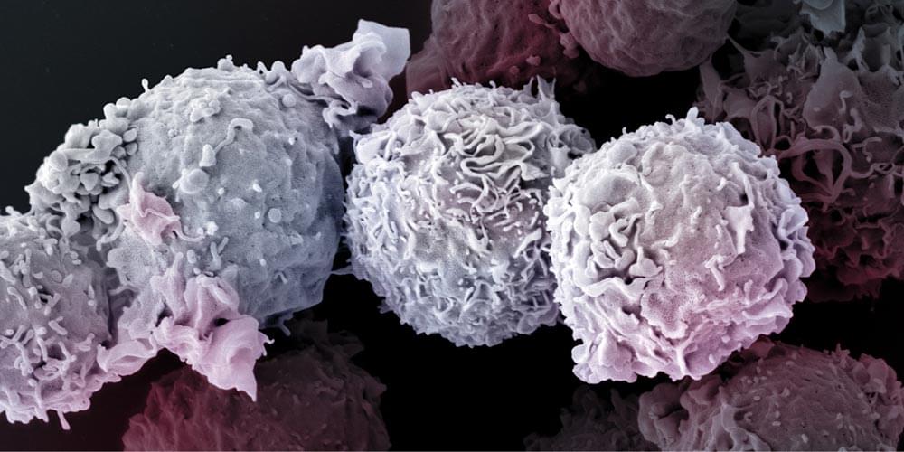
They are at the forefront in the fight against viruses, bacteria, and malignant cells: the T cells of our immune system. But the older we get, the fewer of them our body produces. Thus, how long we remain healthy also depends on how long the T cells survive. Researchers at the University of Basel have now uncovered a previously unknown signaling pathway essential for T cell viability.
Like human beings, every cell in our body tries to ward off death as long as it can. This is particular true for a specific type of immune cells, called T-lymphocytes, or T cells for short. These cells keep viruses, bacteria, parasites and cancerous cells at bay. While T cell production is an active process in infants, children and young adults, it comes to a gradual stop upon aging, meaning that in order to maintain adequate immunity up to an old age, your T cells should better live as long as you.
How T cells manage to survive for such a long time, up to several decades in humans, has long remained unclear. In collaboration with scientists at the Department of Biomedicine and sciCORE, the Center for Scientific Computing of the University of Basel, Professor Jean Pieters’ research group at the Biozentrum has now revealed the existence of a hitherto unrecognized pathway promoting long-term survival of T cells. In Science Signaling they report that this signaling pathway, regulated by the protein coronin 1, is responsible for suppressing T cell death.
A good deal of evidence points to declining kidney function as a cause of declining cognitive function in aging. There are strong correlations between loss of kidney function and risk of dementia, for example. Correlation isn’t a smoking gun in matters of aging, however: it is possible for any one of the underlying forms of molecular damage that cause aging, or for intermediate consequences of that damage, to give rise to otherwise unrelated pathologies in different parts of the body. Those pathologies appear more often in people with greater amounts of that form of damage, and thus appear correlated.
Nonetheless, there are good reasons to think that kidney failure and its downstream consequences contribute meaningful to neurodegeneration, perhaps largely by degrading the function of the vascular system. Vascular aging can cause damage and dysfunction in brain tissue via numerous mechanisms, including the pressure damage of hypertension, similar damage resulting from an acceleration of atherosclerosis, failing to delivery sufficient nutrients and oxygen to the energy-hungry brain, and disruption of the blood-brain barrier, allowing inflammatory cells and molecules into the brain.