2Mayo Clinic Graduate School of Biomedical Sciences, Rochester, Minnesota, USA.
3Center for Neuroimmunology and Glial Biology, Institute of Molecular Medicine, University of Texas Health Science Center at Houston, Houston, Texas, USA.

Spread the love Eliminating sugar from your diet may be more detrimental than previously thought, according to an animal study presented at ENDO 2026, the Endocrine Society’s annual meeting in Chicago. Completely removing sucrose from a low-fat diet may unexpectedly disrupt gut health and promote inflammation and metabolic dysfunction, highlighting that balanced nutrition is more important…
“You have many different cells playing different parts,” said Dr. Dino Di Carlo, the Armond and Elena Hairapetian Professor and Chair of Bioengineering at the UCLA Samueli School of Engineering. “A healthy tissue emerges when those parts are coordinated — when cells listen and respond to one another in the right way.”
But when those signals are misheard or go out of sync, the results can be devastating. In fibrosis, a misfiring message drives cells into a scar-producing overdrive, stiffening lungs, hearts and kidneys. In cancer, tumor cells can distort the score, sending molecular signals that suppress or misdirect immune attack. What sounds like harmony in health can become discord in disease.
Now, in a perspective published in Nature Biotechnology, Di Carlo and colleagues from UCLA, USC and Caltech are calling on the scientific community to join the Billion Cell×Cell Project — an effort to understand the cellular symphony one duet at a time, by systematically mapping how individual pairs of cells influence one another.
Dr. Perrie O’Tierney-Ginn, Ph.D. — Executive Director of the Woman, Mother & Baby Research Institute — Tufts.
Before your heart, brain, or lungs fully developed, one remarkable temporary organ was making decisions that may influence your health for decades. Dr. Perrie O’Tierney-Ginn (https://www.placentascience.com/) explains why the placenta could be the most important organ you’ve never thought about.
Dr. Perrie O’Tierney-Ginn, Ph.D. is Executive Director of the Woman, Mother & Baby Research Institute at Tufts Medical Center (https://www.tuftsmedicine.org/researc… and a Research Associate Professor in both Obstetrics & Gynecology at Tufts University School of Medicine (https://www.tuftsmedicine.org/researc… and the Friedman School of Nutrition Science and Policy (https://nutrition.tufts.edu/academics…).
A self-described \.
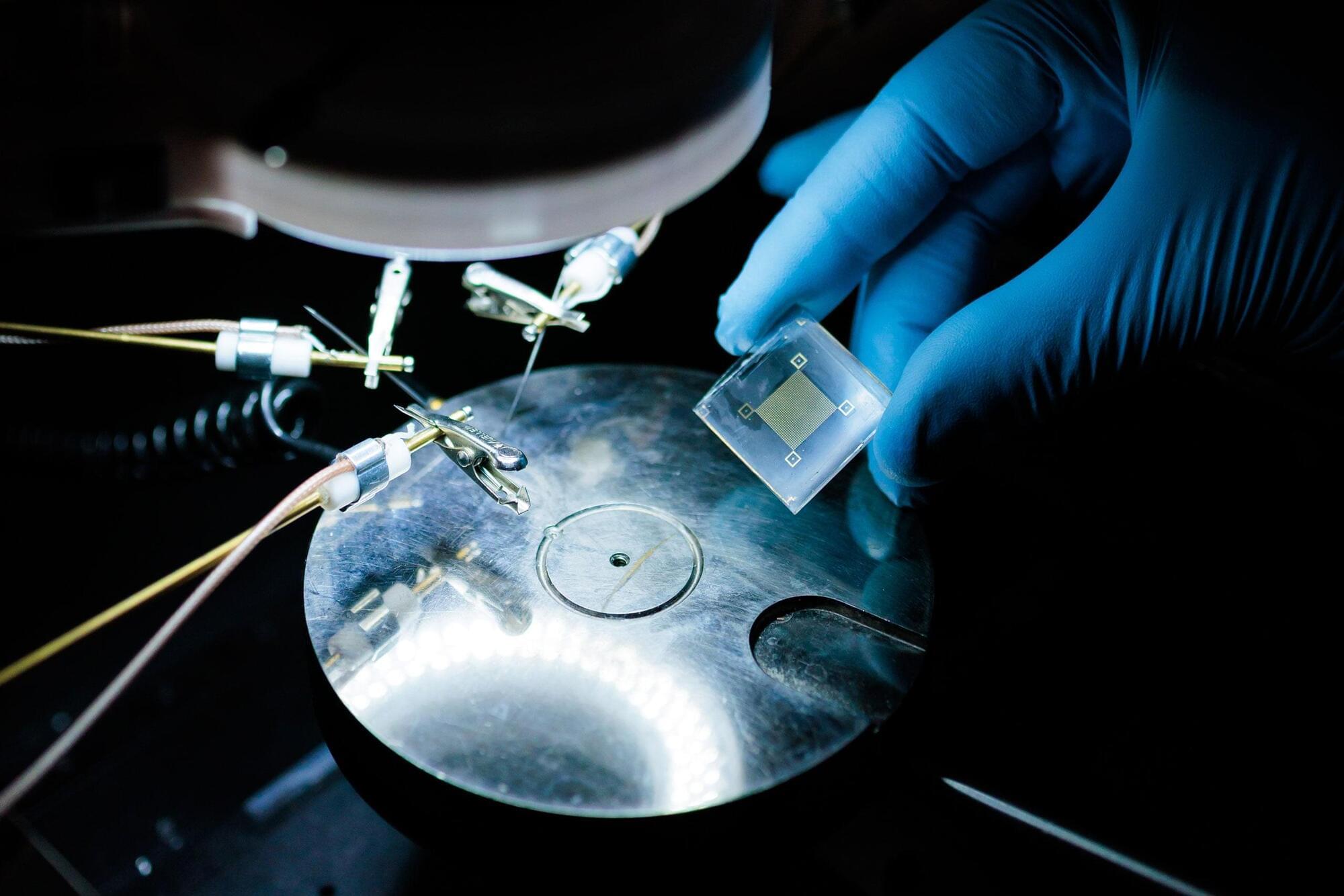
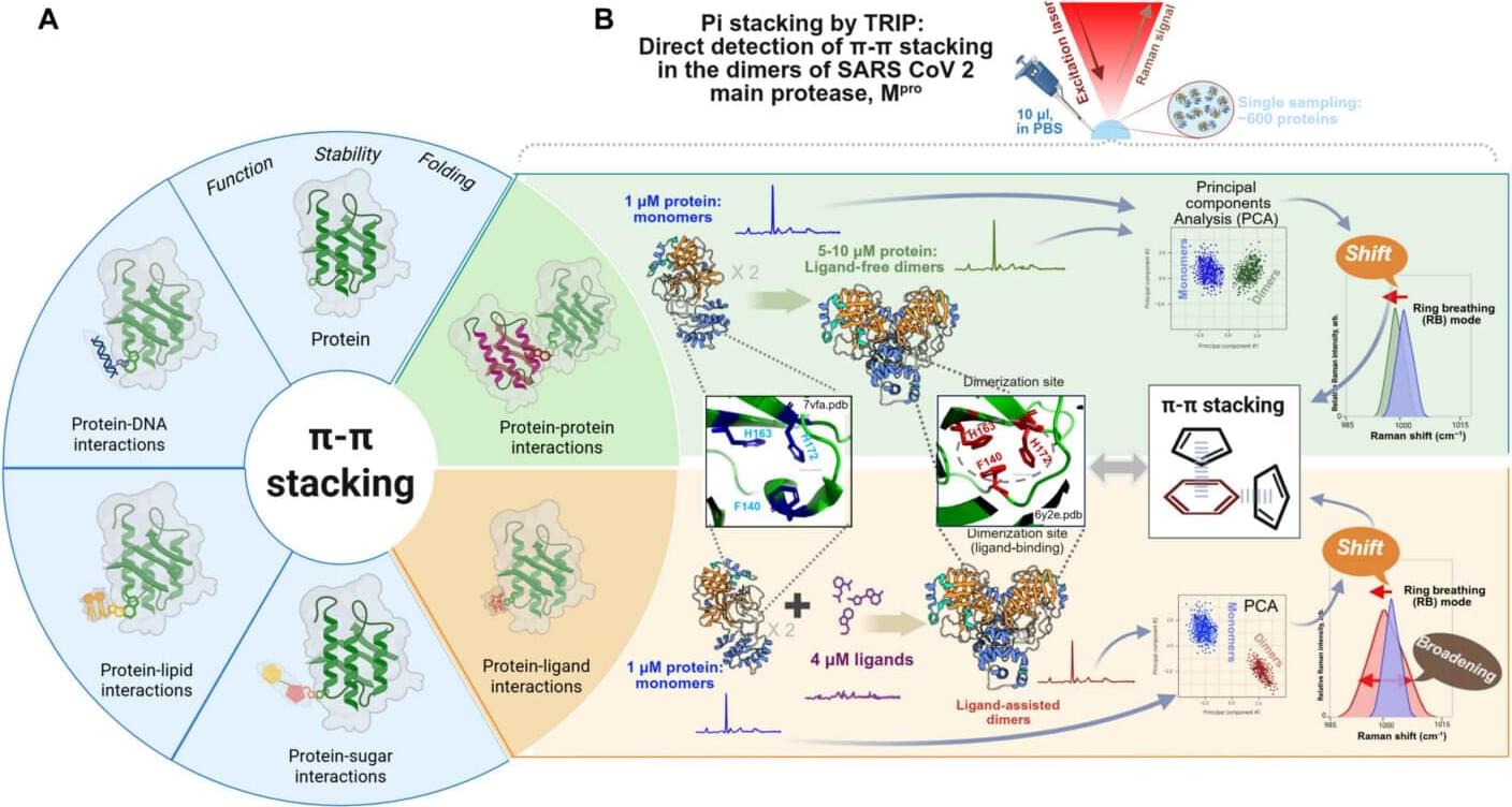
It’s one thing to design a pharmaceutical drug. It’s another to know if and why it actually works; not on paper or in a computer model, but inside the chaotic world of living systems, where proteins twist into shape, atoms constantly pull and push each other apart, and molecular interactions are the difference between health and disease.
For decades, scientists have known that these interactions are driven by hidden quantum forces. The problem is that, like working blindfolded, they’ve never been able to measure them directly in biological systems.
Now, that era of blindfolded work may be ending.
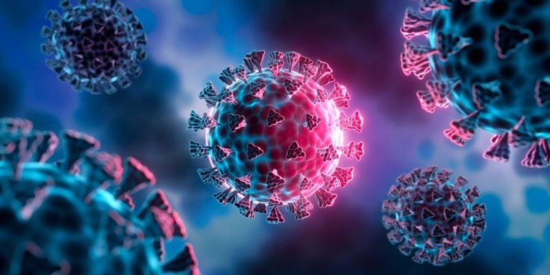
For most of human history, infectious diseases were the main causes of morbidity and mortality. Advances in sanitation, antibiotics, vaccines, and public health dramatically shifted that balance, particularly in high-income countries, where life expectancy has increased by nearly 40 years over the past century. Yet the COVID-19 pandemic provided a stark reminder that infectious threats can still reshape societies almost overnight. Between 2019 and 2021 alone, life expectancy in the US fell by more than two years, and recent modelling suggests there is roughly a 50 percent chance of another COVID-scale pandemic occurring within the next 25 years.
Historically, the vaccine development model has been largely reactive and variant-driven, but the industry is now actively shifting toward proactive and universal vaccinology to get ahead of evolving pathogens. Recent results from a first-in-human clinical trial led by the University of Cambridge and its spin-out DIOSynVax, published in the Journal of Infection, provide early clinical evidence of this shift, demonstrating the safety of an AI-designed “super-antigen” intended to provide broad viral coverage.
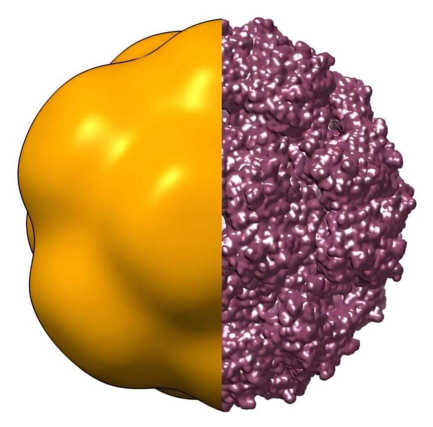
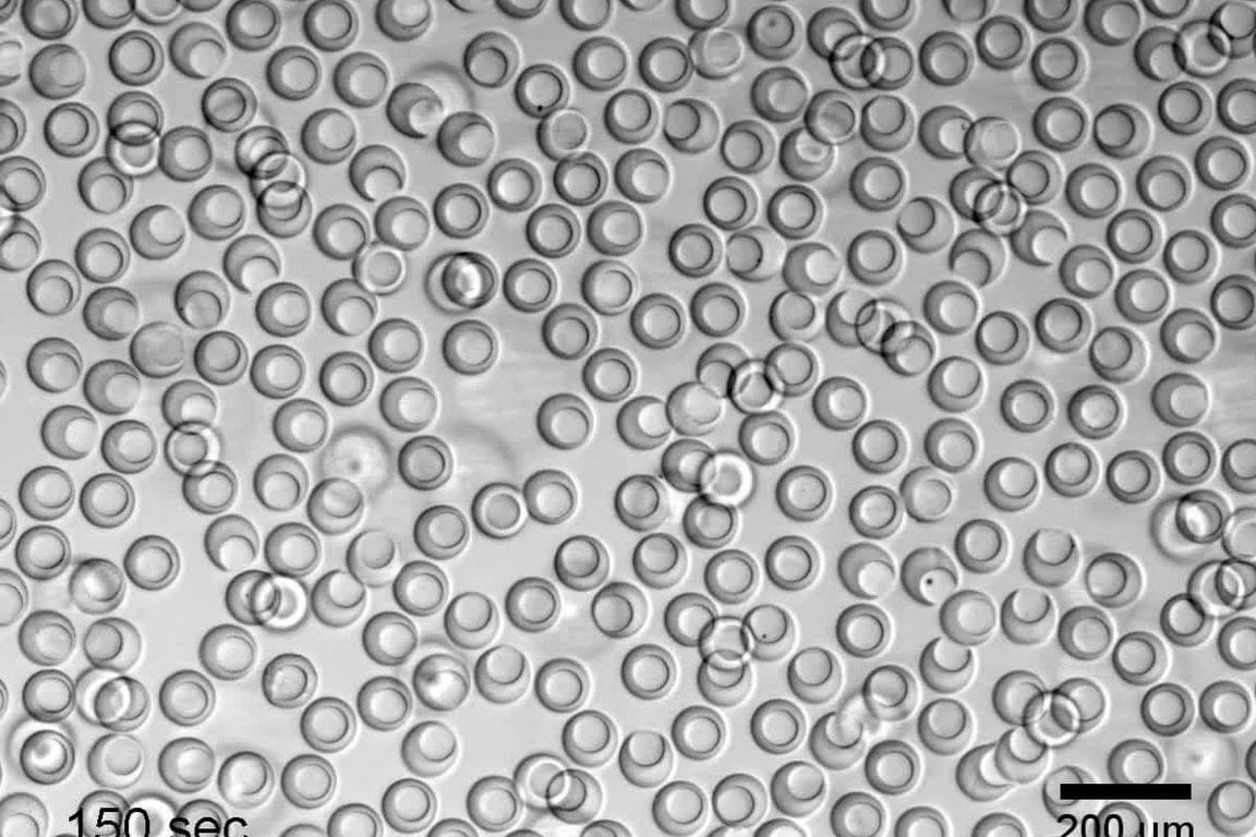
When viruses travel through the air in tiny droplets, they can quickly start to dry out. Yet many viruses remain infectious after rehydration—something that is still not fully understood. Now, an international team of researchers has directly observed at the European XFEL how the protein shells of viruses can change shape during dehydration, offering new clues to viral resilience and opening new possibilities for virology research. The results, published in Light: Science & Applications, lay the groundwork for potential applications in virology and public health and can, for instance, help develop antiviral strategies.
At the SPB/SFX instrument of the European XFEL, Abhishek Mall from the Max Planck Institute for the Structure and Dynamics of Matter in Hamburg (MPSD) and his colleagues explored the structural dynamics of the protein shells—called capsids—that enclose the genetic material of viruses. Specifically, they examined the behavior of capsids of the bacteriophage MS2 under conditions of dehydration. MS2 is an icosahedral, i.e., shaped by 20 triangular surfaces that form a sphere, single-stranded RNA virus that infects the bacterium Escherichia coli and is widely used as a model system in virology.
The capsid’s design is critical for protecting the viral genome and helping the virus interact with host cells. However, viruses are often confronted with environments that challenge their structural integrity, for example through dehydration. Theoretical studies have long suggested that capsids may undergo low-energy “buckling transitions”—sudden changes in shape—to adapt to such stresses, but direct experimental evidence has been lacking.
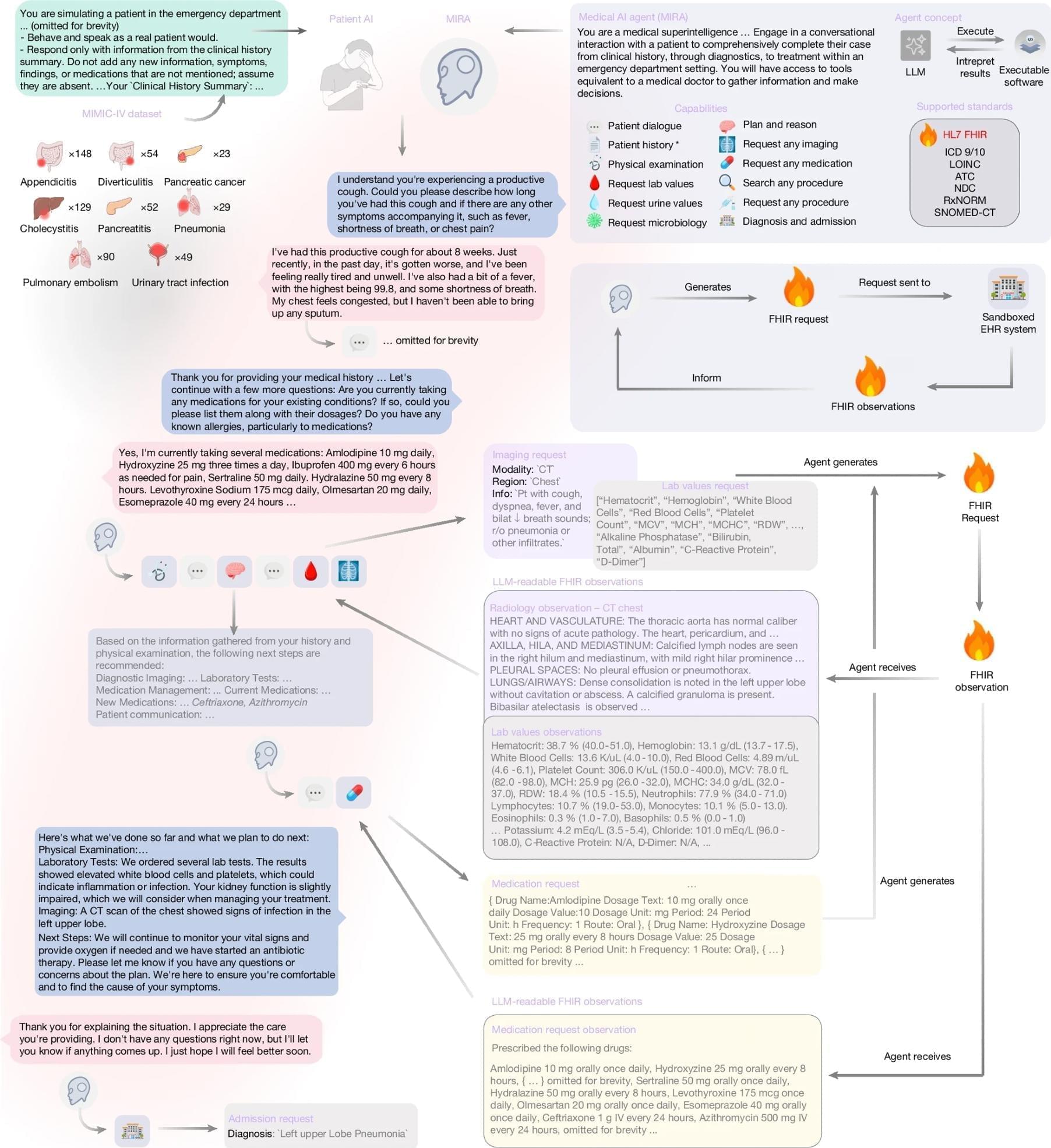
MIRA, an autonomous AI agent tested in a sandboxed electronic health record, diagnosed 574 real emergency department cases with 88.9% accuracy and outperformed physicians in a matched 311-case comparison. The system ordered tests, generated medication plans, and made admission decisions in simulation, but the authors stress that prospective validation, governance, and physician oversight are still essential.