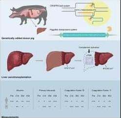
The advent of genetically edited porcine-to-human xenotransplantation has predominantly focused on cardiac and renal applications, with no reported cases of porcine-to-human liver xenotransplantation. This study presents the world’s first successful genetically modified pig auxiliary liver xenotransplantation in a living human, achieving an unprecedented survival of 171 days, and provides valuable insights into the critical factors influencing the procedure’s success.
Category: genetics – Page 91

Genetically encoded biosensor tracks plants’ immune hormone in real time
From willow bark remedies to aspirin tablets, salicylic acid has long been part of human health. It also lies at the heart of how plants fight disease. Now, researchers at the University of Cambridge have developed a pioneering biosensor that allows scientists to watch, for the first time, how plants deploy this critical immune hormone in their battle against pathogens.
Published in Science, Dr. Alexander Jones’s group at the Sainsbury Laboratory, Cambridge University (SLCU) presents SalicS1, a genetically encoded biosensor that can detect and track the dynamics of the plant immune hormone salicylic acid (SA) with exquisite precision inside living plants.
Salicylic acid is a central regulator of plant immunity, triggering defense responses against a huge diversity of invaders. Until now, however, scientists have lacked the tools to measure SA at high enough spatial and temporal resolution to understand how plants balance growth with immune defense.
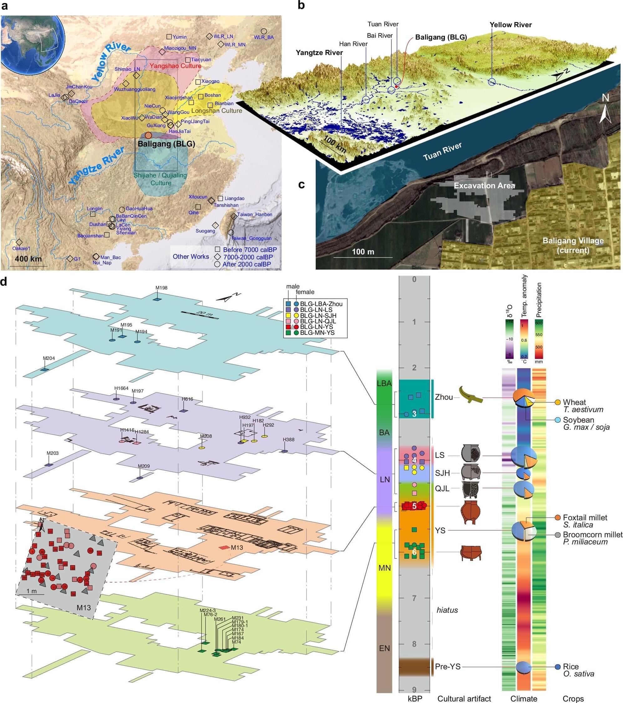
Ancient DNA reveals prehistoric connections and a patrilineal society in early China
Scientists from Peking University have uncovered new genetic evidence that sheds light on how prehistoric people in China interacted, migrated, and built their communities. Led by Professors Huang Yanyi and Pang Yuhong from the Biomedical Pioneering Innovation Center (BIOPIC), the research reveals the first direct genetic proof of a patrilineal social system in Neolithic China.
The study, conducted in collaboration with Yunnan University and Minzu University of China, was published in Nature Communications on September 30, 2025.
The story of Chinese civilization begins along two great rivers: the Yellow River, known for its millet-farming cultures, and the Yangtze River, home to early rice agriculture. How people in these regions exchanged ideas, adapted to environmental shifts, and shaped early societies has long fascinated archaeologists and historians.
Scientists fix genetic defect in mice tied to brain disorders that include autism and epilepsy
In an exciting scientific first, researchers at the Allen Institute successfully designed a new gene therapy that reversed symptoms related to SYNGAP1-related disorders (SRD) in mice. These are a class of brain disorders that can lead to severe and debilitating symptoms including intellectual disability, epilepsy, motor problems, and risk-taking behaviors in humans. In most cases, SRDs are caused when someone has only one working copy of the SYNGAP1 gene instead of the normal two.
The findings, recently published in the journal Molecular Therapy, represent the first successful gene supplementation therapy for SRDs in which an adeno associate virus (AAV) was used to deliver a working copy of the SYNGAP1 gene into brain cells. AAVs are non-replicating viruses that act like delivery trucks carrying therapeutic cargo, in this case the SYNGAP1 gene, into cells that need it.
“Gene supplementation is providing a functional new copy of a defective gene, a strategy that has great potential for correcting diseases where a gene is completely missing or where a single copy of a gene is lost,” said Boaz Levi, Ph.D., associate investigator at the Allen Institute and senior author of the study. “This provides a clear demonstration that SYNGAP1-related disorders can be treated with a neuron-specific gene supplementation strategy. It’s an important milestone for the field that provides hope for those who suffer from this class of severe neurological diseases.”

Synaptic Dysfunction in Dementia Can Be Modelled in Patient-Derived Neurons
Neurons produced from frontotemporal dementia patients’ skin biopsies using modern stem cell technology recapitulate the synaptic loss and dysfunction detected in the patients’ brains, a new study from the University of Eastern Finland shows.
Frontotemporal dementia is a progressive neurodegenerative disease affecting the frontal and temporal lobes of the brain. The most common symptoms are behavioral changes, difficulties in understanding or producing speech, problems in movement, and psychiatric symptoms. Often, frontotemporal dementia has no identified genetic cause, but especially in Finnish patients, hexanucleotide repeat expansion in the C9orf72 gene is a common genetic cause, present in about half of the familial cases and in 20 per cent of the sporadic cases where there is no family history of the disease. However, the disease mechanisms of the different forms of frontotemporal dementia are still poorly understood, and there are currently no effective diagnostic tests or treatments affecting the progression of the disease in clinical use.
Brain imaging and neurophysiological studies have shown that pathological and functional changes underlying the symptoms occur at synapses, the connections between brain neurons, in frontotemporal dementia patients. PET imaging studies have shown significant synapse loss in the brain, and transcranial magnetic stimulation, on the other hand, has indicated disturbed function of both excitatory and inhibitory neurotransmitter systems, leading to deficient neurotransmission. Often, drugs affecting the different neurotransmitter systems are used to mitigate the symptoms of frontotemporal dementia patients.
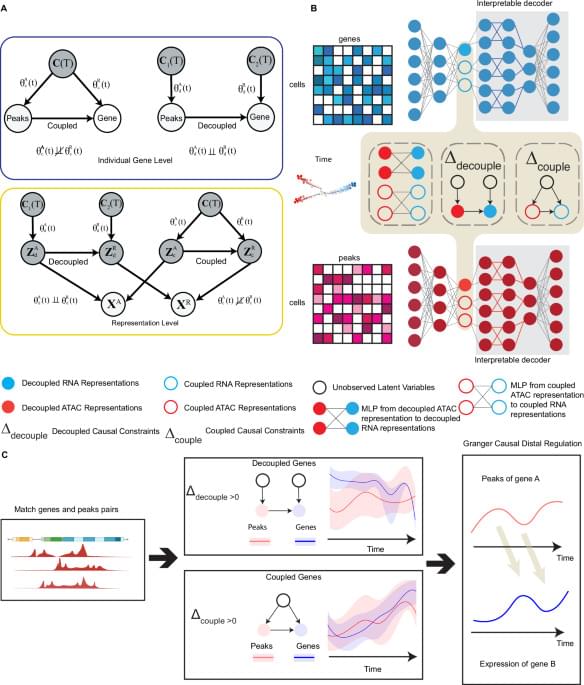
HALO: hierarchical causal modeling for single cell multi-omics data
Chromatin accessibility dynamics causally influence changes in gene expression levels, but these fluctuations may not be directly coupled over time. Here, authors develop computational causal framework HALO, examining epigenetic plasticity and gene regulation dynamics in single-cell multi-omic data.

Alzheimer’s disease research in brain tissue from African American donors points to roles for many novel genes
The prevalence of Alzheimer’s disease (AD) is approximately two times higher in African Americans (AA) compared to white/European-ancestry (EA) individuals living in the U.S. Some of this is due to social determinants of health such as disparities in health care access and quality of education, biases in testing and higher rates of AD risk factors such as cardiovascular disease and diabetes in those who identify as African American.
Although many studies have examined differences in gene expression (a measure of the amount of protein encoded by a gene) in brain tissue from AD cases and controls in EA or mixed ancestry cohorts, the number of AA individuals in these studies was unspecified or too small to identify significant findings within this group alone.
In the largest AD study conducted in brain tissue from AA donors, researchers from Boston University Chobanian & Avedisian School of Medicine have identified many genes, a large portion of which had not previously been implicated in AD by other genetic studies, to be significantly more or less active in tissue from AD cases compared to controls. The most notable finding was a 1.5 fold higher level of expression of the ADAMTS2 gene in brain tissue from those with autopsy-confirmed AD.
Depression genetics differ by sex: Study find females carry higher risk than males do
Important genetic differences in how females and males experience depression have been revealed for the first time in findings that could pave the way for more targeted intervention and treatments.
In the study, published in Nature Communications, scientists found that genetic factors contribute more to depression risk in females than in males. The team discovered about twice as many genetic “flags” for depression in the DNA of females as they did in that of males.
“We already know that females are twice as likely to suffer from depression in their lifetime than males,” said Dr. Brittany Mitchell, Senior Researcher at QIMR Berghofer’s Genetic Epidemiology Lab. “And we also know that depression looks very different from one person to another. Until now, there hasn’t been much consistent research to explain why depression affects females and males differently, including the possible role of genetics.”