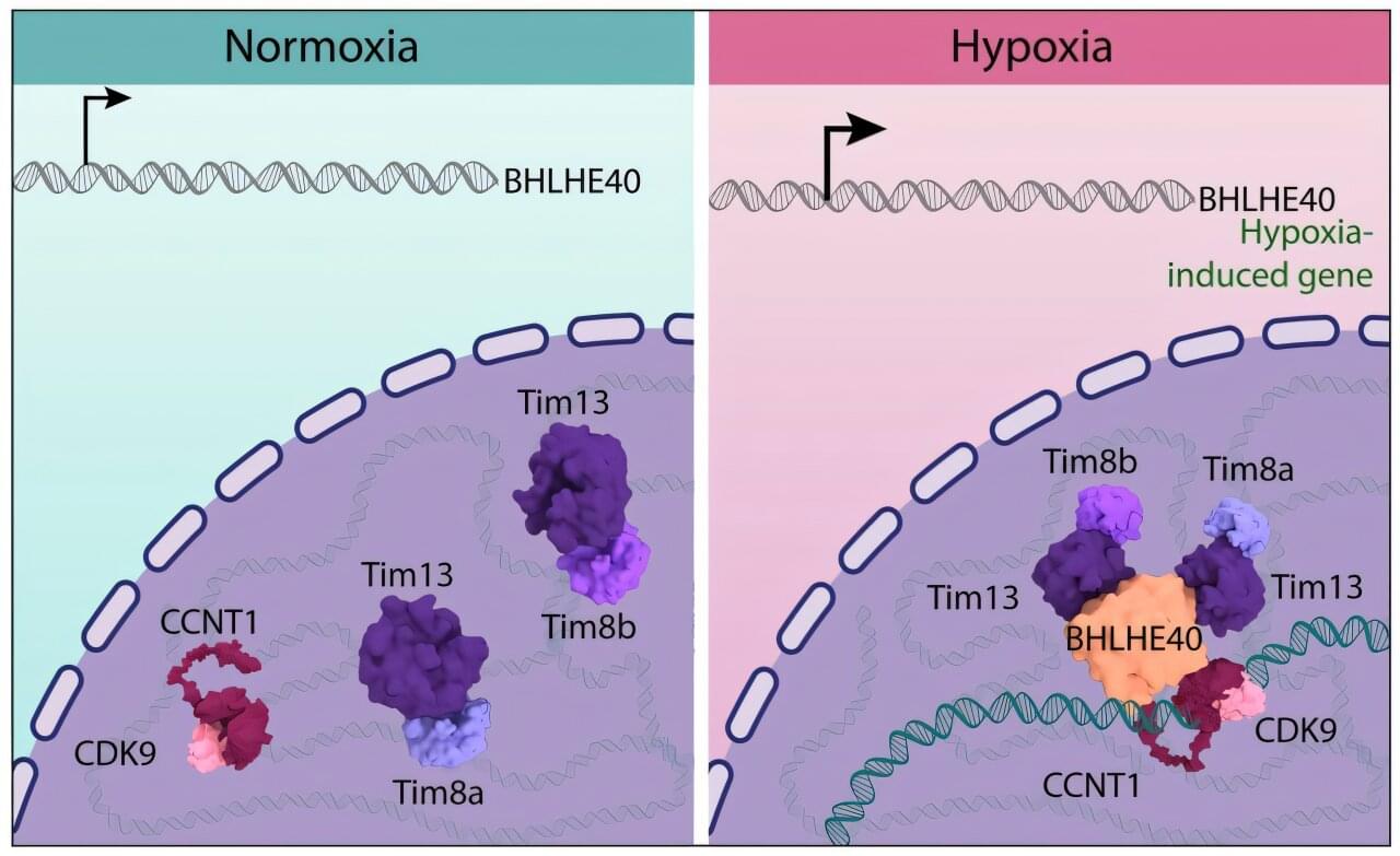
Northwestern Medicine scientists have, for the first time, described the underlying mechanisms that regulate how cells rapidly change gene expression in response to hypoxia, a key feature of many treatment-resistant tumors, according to a recent study published in Science Advances.
Ali Shilatifard, Ph.D., the chair and Robert Francis Furchgott Professor of Biochemistry and Molecular Genetics, was the senior author of the study.
Scientists have uncovered the oldest human genetic code from an 8-year-old Neanderthal child in Belgium, offering profound insights into our evolutionary past and Neanderthal development.
A new study from the University of Bergen shows an association between breastfeeding up to 6 months of age and a reduced risk of ADHD symptoms from ages 3 to 8.
Breast milk is the primary source of nutrition for infants. It is uniquely tailored for the child and contains numerous components beneficial for growth and brain development, including long-chain fatty acids, amino acids, antibodies and beneficial bacteria.
“It is well established that psychiatric symptoms and disorders can be influenced by both genetic and environmental factors,” says Berit Skretting Solberg, psychiatrist and researcher at the Department of Biomedicine, University of Bergen, and senior consultant at Betanien Hospital.
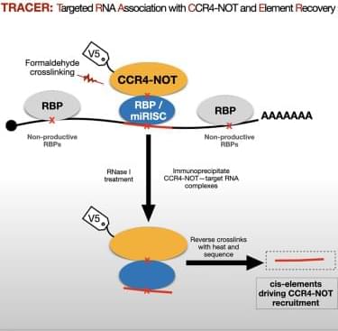
Luo et al. present TRACER, a transcriptome-wide approach to identify RNA elements that recruit the CCR4-NOT complex. TRACER uncovers thousands of CCR4-NOT-associated elements, many mapping to known or predicted RBP and miRNA target sites. These elements drive mRNA repression and can be targeted using gene editing or ASO approaches.
Life Biosciences just dosed the first human patient with ER-100 — an OSK gene therapy built from three Yamanaka factors, designed to reprogram old cells back toward a younger state. The first target is the eye. But the real implication is much bigger: this method appears to work on every tissue type it has been tried on.
If this first eye trial comes back safe, it could be the first domino in a much larger age-reversal wave: eye, liver, brain, skin, muscle, heart, kidney, blood vessels — potentially every tissue in the body.
This episode reveals the tidal wave of companies racing toward human trials using the same basic strategy: epigenetically reprogramming old cells so they behave young again. Billions of dollars are pouring into this from Jeff Bezos, Sam Altman, Brian Armstrong, Peter Thiel, and the biggest names in longevity biotech.
We walk through who they are, what they are trying to cure, why the eye came first, what worked in mice and monkeys, why NewLimit is going after liver rejuvenation, and whether the cheap pill version could be right behind the expensive gene therapy.
Bottom line: real age reversal is now in a human trial.
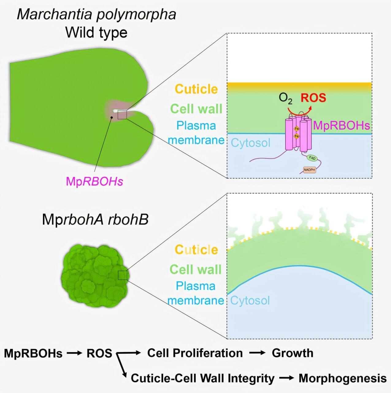
Reactive oxygen species (ROS) produced naturally during cellular metabolism often cause oxidative damage to cells. However, these molecules also play an important role in normal cellular signaling. While ROS are established as essential signaling molecules in various organisms, their precise role in basic plant development and morphogenesis remains unclear.
A family of enzymes known as NADPH oxidases (NOXs) generates ROS that act as physiologically important signaling molecules. In plants, the NOX enzymes are known as respiratory burst oxidase homologs (RBOHs), which are implicated in diverse physiological processes. However, their contribution to plant development, including cell proliferation and ordered morphogenesis, has remained insufficiently understood.
To address this gap, a team of researchers led by Professor Kazuyuki Kuchitsu from the Department of Applied Biological Science at Tokyo University of Science (TUS) in Japan conducted a study.
Researchers stimulated human neurons and tracked gene activity. Activation exposed hidden variants and epigenetic changes linked to schizophrenia and autism, explaining disease risk missed in resting brain studies.
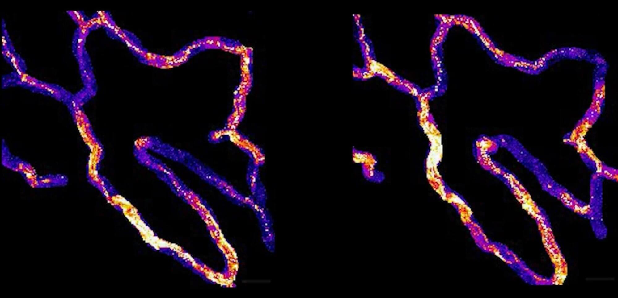
The cells lining skin capillaries are constantly sending each other messages—tiny pulses of calcium that help regulate blood flow, sense physical forces and keep vessel walls intact. Scientists have known about this signaling for decades. What they didn’t know, until now, is that it follows a remarkably organized pattern, one that persists across days and weeks, governed by a network of cells that have, in a sense, assigned themselves permanent roles.
A new study from Yale School of Medicine (YSM) and University of California, Los Angeles (UCLA), published in Proceedings of the National Academy of Sciences, reveals not only that this network exists, but also what happens when it breaks down—and how it might be restored.
The study was done in the lab of Valentina Greco, Ph.D., Carolyn Walch Slayman Professor of Genetics at YSM and a Howard Hughes Medical Institute investigator, in close collaboration with the labs of Julia Mack, Ph.D., and Chen Yuan Kam, Ph.D., both at UCLA.
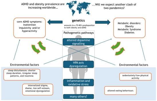
Attention-deficit/hyperactivity disorder (ADHD) is a neurodevelopmental disorder characterized by inattention, impulsivity and/or hyperactivity. In recent years, metabolic alterations, primarily obesity, insulin resistance, and diabetes, have emerged as frequent comorbidities in individuals with ADHD, suggesting a bidirectional relationship between neurodevelopmental and metabolic dysfunctions. Emerging evidence indicates that dysregulation of dopaminergic signaling, disturbances in the hypothalamic-pituitary-adrenal (HPA) axis, and chronic low-grade inflammation are central to both ADHD symptomatology and metabolic impairments. For instance, alterations in dopamine-related genes (e.g., DRD4, DAT1) not only affect cognitive and behavioral functions but also play a role in appetite regulation and glucose homeostasis. Epidemiological studies further demonstrate that individuals with ADHD exhibit poorer glycemic control and a higher prevalence of both type 1 and type 2 diabetes, while early-life metabolic challenges such as maternal diabetes may predispose offspring to ADHD. This review aims to comprehensively synthesize the epidemiological, genetic, and pathogenetic evidence linking ADHD to metabolic alterations. We discuss key pathophysiological pathways—including dopaminergic dysregulation, HPA axis disturbances, inflammation, and oxidative stress—and evaluate their contributions to the co-occurrence of ADHD and metabolic disorders. In addition, we explore the clinical implications and integrated treatment approaches that encompass lifestyle modifications, pharmacological therapies, and multidisciplinary care. Finally, we outline future research directions to develop personalized and holistic interventions.


