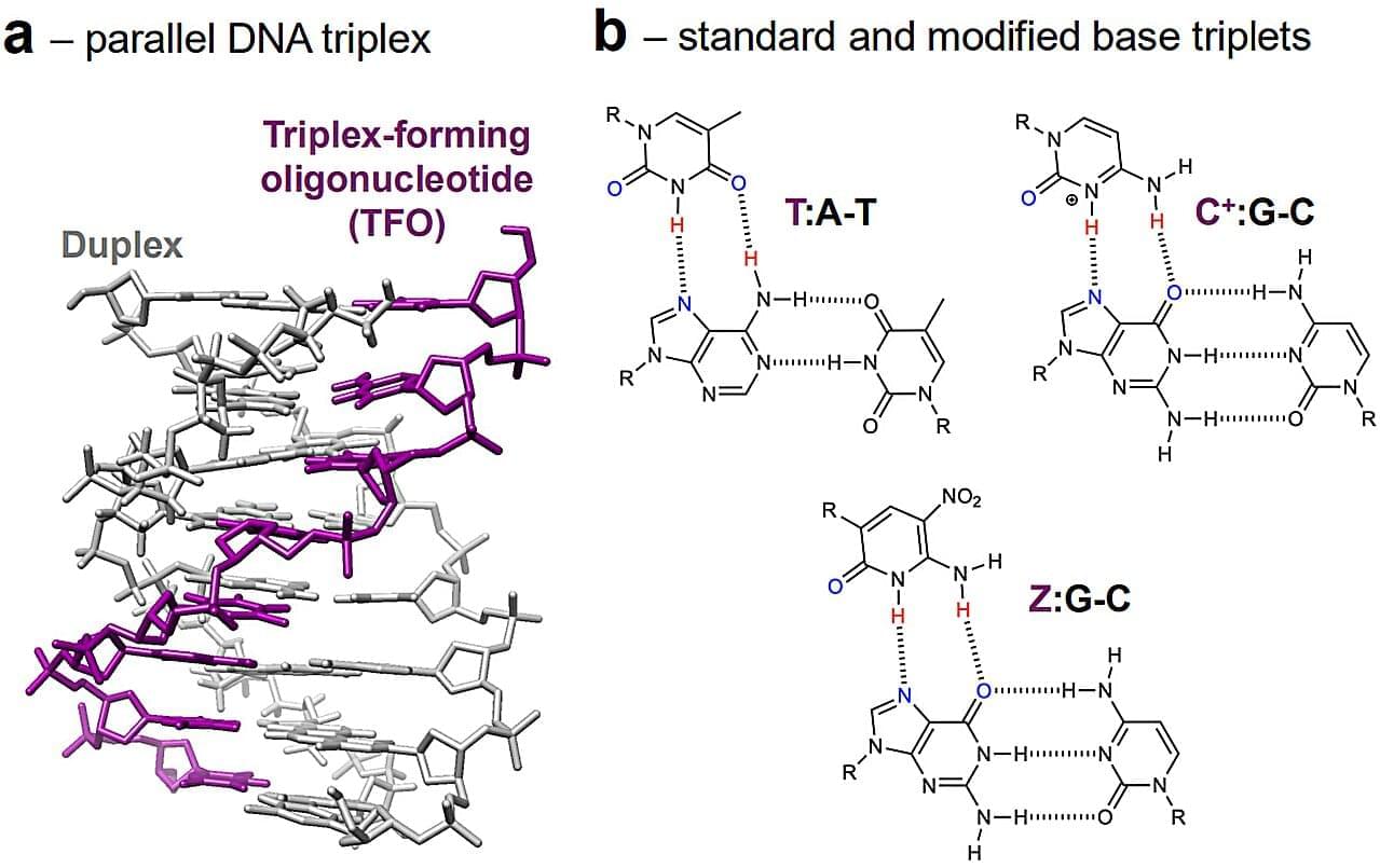
Toggle light / dark theme

Specialized transporters relay lipids to cellular targets

In addition to providing energy, lipids are also essential building blocks of our cell membranes. However, despite their importance, they remain poorly understood. A research team has revealed for the first time the secrets of their transport within cells. Each lipid uses a limited number of proteins to move from its place of production to its place of action. The team has also compiled an inventory of the proteins involved in the transport of hundreds of lipids.

These findings, published in the journal Nature, provide a better picture of the functioning of our cells, as well as of many genetic and metabolic disorders, such as diabetes and Alzheimer’s disease.

Biologists brought together more than a hundred transfer proteins with hundreds of different lipids. The aim was to obtain the most comprehensive list possible of the ‘pairs’ formed between each protein and the lipids it can carry.

To do this, two experimental methods were combined. The first, carried out in a test tube, provides a highly controlled environment, while the second, which more closely corresponds to the inside of a cell, allows researchers to verify how these bonds are formed under near-real conditions. This is a world first on such a scale and at such a level of complexity. “The ‘‘couples’’ identified show that transfer proteins are not “buses” capable of transporting most lipids, but private chauffeurs with specific characteristics,” explains the senior author.

Scientists have been able to determine, using advanced mathematical models, how three transfer proteins recognise, among all lipids, those that they actually transport. ScienceMission sciencenewshighlights.

Scientists identify promising new target for Alzheimer’s-linked brain inflammation

A multidisciplinary team has developed a selective compound that inhibits an enzyme tied to inflammation in people at genetic risk for Alzheimer’s, while preserving normal brain function and crossing the blood-brain barrier.

The findings are published in the journal npj Drug Discovery.

The driver is an enzyme called calcium-dependent phospholipase A2 (cPLA2). The team discovered its role in brain inflammation by studying people who carry the APOE4 gene —the strongest genetic risk factor for Alzheimer’s disease. While many people who have the APOE4 gene don’t develop the disease, those with elevated levels of cPLA2 generally do.

How beige fat keeps blood pressure in check

In this report, researchers link thermogenic adipose tissue (brown/beige fat), best known for heat production, to blood-pressure control via direct fat–blood vessel communication. Using mouse models engineered to lose beige fat identity (via adipocyte-specific disruption of PRDM16), they observed elevated arterial pressure alongside perivascular remodeling, including fibrotic tissue accumulation and marked vascular hypersensitivity to the vasoconstrictor hormone angiotensin II. Mechanistically, loss of beige fat identity increased secretion of QSOX1 (quiescin sulfhydryl oxidase 1), which activated pro-fibrotic gene programs in vascular cells and promoted vessel stiffening; blocking this pathway (including genetic removal of QSOX1 in the model) restored healthier vascular signaling and function. The authors characterize this as a previously underappreciated, obesity-independent axis by which the “quality” (thermogenic vs white-like) of perivascular fat influences vascular stiffness and responsiveness to pressor signals, suggesting QSOX1 and related adipose-derived signals as potential precision targets for future antihypertensive therapies.


A mouse aorta with immunofluorescent tagging, emphasizing the close connection between vasculature and fat. (Credit: Cohen lab)

Obesity causes hypertension. Hypertension causes cardiovascular disease. And cardiovascular disease is the leading cause of death worldwide. While the link between fat and high blood pressure is clearly central to this deadly chain, its biological basis long remained unclear. What is it about fat that impacts vascular function and blood pressure control?

Now, a new study demonstrates how thermogenic beige fat—a type of adipose tissue, distinct from white fat, that helps the body burn energy—directly shapes blood pressure control. Building on clinical evidence that people with brown fat have lower odds of hypertension, the researchers created mouse models that cannot form beige fat (the thermogenic fat depot in mice that most closely resembles adult human brown fat) to watch what happens when this tissue is lost. They found that the loss of beige fat increases the sensitivity of blood vessels to one of the most important vasoconstricting hormones (angiotensin II)—and that blocking an enzyme involved in stiffening vessels and disrupting normal signaling can restore healthy vascular function in mice. These results, published in Science (opens in new window), reveal a previously unknown mechanism driving high blood pressure and point toward more precise therapies that target communication between fat and blood vessels.

Elite army training reveals genetic markers for resilience

A new analysis of soldiers attempting to join the U.S. Army Special Forces suggests that specific genetic variations play a role in how individuals handle extreme physical and mental pressure. The research identified distinct links between a soldier’s DNA and their cognitive performance, psychological resilience, and physiological stress response during a grueling selection course. These findings were published recently in the academic journal Physiology & Behavior.

To become a member of the elite Army Special Forces, a soldier must first pass the Special Forces Assessment and Selection course. This training program is widely recognized as one of the most difficult military evaluations in the world. Candidates must endure nearly three weeks of intense physical exertion. They face sleep deprivation and complex problem-solving exercises. The attrition rate is notoriously high. Approximately 70 percent of the soldiers who attempt the course fail to complete it. This environment creates a unique laboratory for scientists to study human endurance.

Researchers have sought to understand why some individuals thrive in these punishing environments while others struggle. Resilience is generally defined as the ability to adapt positively to adversity, trauma, or threats. It involves a combination of psychological stability and physiological recovery. While physical training and mental preparation are essential, biological factors also play a substantial role. Genetics help determine how the brain regulates chemicals and how the body processes stress hormones.

Intestinal epithelial TLR5 signaling promotes barrier-supportive macrophages

Crosstalk between commensal E. coli that express flagellin and intestinal epithelial cells coordinate intestinal macrophage recruitment to support gut barrier homeostasis in mice.

Learn more in ScienceImmunology.


The colonic epithelium is an important boundary between internal tissues and luminal contents including the microbiota. The gut microbiota drives immune cell accumulation and effector function (6, 10, 12), but how colonic epithelial cells mediate these processes is incompletely understood. To understand how intestinal epithelial sensing of adherent microbes regulates immune-supported intestinal barrier repair, we used the E. coli strain 541–15, which we previously found increased LP macrophages and promoted their IL-10 production, protecting against inflammatory pathology in mouse colitis models (12, 23, 24).

Here, we demonstrated that E. coli 541–15 colonization promotes LP recruitment of mature macrophages after antibiotic treatment. Using HCMs, we determined that E. coli 541–15 induced expression of immune regulatory genes including the monocyte-recruiting chemokine CCL2 exclusively in UD cells, which promote monocyte migration. In vivo, CCL2 produced by epithelial cells in response to E. coli 541–15 colonization promoted colon LP macrophage expansion and protected mice from DSS colitis. We further identified flagellin as the key microbial factor that induced epithelial CCL2 expression. Last, epithelial TLR5 and E. coli flagellin were both required for LP recruitment of mature macrophages and protection against DSS challenge. In both in vitro and in vivo systems, epithelial stem cells had higher TLR5 expression than mature IECs, indicating a crypt specific role for flagellated bacteria detection. Our findings are consistent with previous studies showing that TLR5-deficient mice develop spontaneous colitis in the presence of the pathobiont H. hepaticus (20), suggesting a potential protective role for TLR5 in intestinal homeostasis. Moreover, other studies report that H. hepaticus induces colitis in IL-10–deficient mice (52, 53), highlighting a possible link between TLR5 and IL-10+ macrophages in H. hepaticus pathogenesis. Here, we demonstrate that TLR5 signaling is essential for mucosal protection by promoting the recruitment of CCR2+ cells and the maturation of LP macrophages, which are key producers of IL-10 in the gut, highlighting a possible link between TLR5 and IL-10+ macrophages in H. hepaticus pathogenesis.

Previous work demonstrated that TLR5 expression differs by intestinal region, with expression restricted to Paneth cells in the small intestine crypt but distributed more broadly among colonic epithelial cells (54). Three-dimensional (3D) mouse Paneth cells enriched small intestinal organoids, and colonoids (which contain both undifferentiated and differentiated cells) responded to flagellin and up-regulated chemokines (54); however, the specific flagellin-responsive colonic cell types remained undefined. In addition, early studies using human epithelial cell lines showed that TLR5 localizes to the IEC basolateral surface, suggesting that flagellin sensing is limited to situations where bacterial products cross the epithelial barrier (55, 56). Our current study advances this understanding by using HCMs that allow for functional separation of DF IECs and UD stem-like cells, which express higher TLR5. After apical or basolateral treatment, UD, but not DF, HCMs responded to TLR5 stimulation. Similar to HCMs, in the mouse epithelium, we found higher Tlr5 expression in LGR5+ stem cells than mature LGR5 IECs. These results suggest that colonic stem cells in humans and mice, such as Paneth cells in the small intestine, act as critical sensors of flagellated microbes and highlight a conserved mechanism to spatially restrict microbial recognition to the crypt base to safeguard the stem cell niche. Under homeostatic conditions, stem cells are physically shielded from microbial stimulation by mucus, secretory immunoglobulin A (IgA), and antimicrobial peptides (13). However, multiple studies showed colonization of cecal and colonic crypts with select flagellated commensal bacteria at homeostasis, which could induce TLR5 signaling (4749, 57). Furthermore, disruption of the epithelial barrier during injury and resulting expansion of the stem cell zone may increase stem cell and microbial interactions. We propose that compartmentalized TLR5 signaling provides a protective strategy, which promotes tonic macrophage expansion in the steady state and enables amplification when epithelial integrity is compromised or after colonization with microbes that can reach the base of the crypt.

A genetic breakthrough links early-onset diabetes to brain disorders

Paediatric teams are now facing babies whose diabetes appears in the first weeks of life, then rapidly reveals deep problems in brain growth and function. A new genetic finding sheds light on how a single molecular fault can disrupt both blood sugar control and early brain development.

Neonatal diabetes is diagnosed in the first six months of life, often within days or weeks after birth. Unlike the more common type 1 diabetes, which usually shows up in children and teenagers, neonatal diabetes is almost always genetic.

Doctors typically notice poor feeding, weight loss, dehydration and extremely high blood sugar. In many cases, the root cause is a mutation that stops the pancreas from making enough insulin. That alone makes neonatal diabetes a medical emergency.

Two harmful gene variants can restore function when combined, study reveals

Sometimes, in genetics, two wrongs do make a right. A research team has recently shown that two harmful genetic variants, when occurring together in a gene, can restore function—proving a decades-old hypothesis originally proposed by Nobel laureate Francis Crick.

Their study, to be published in the Proceedings of the National Academy of Sciences, not only experimentally validated this theory but also introduced a powerful artificial intelligence (AI)-driven approach to genetic interpretation led by George Mason University researchers.

The project began when Aimée Dudley, a geneticist at the Pacific Northwest Research Institute (PNRI), approached George Mason University Chief AI Officer Amarda Shehu after following her lab’s work on frontier AI models for predicting the functional impact of genetic variation. That conversation sparked a collaboration that married PNRI’s experimental expertise with George Mason’s computational innovation to discover some surprising ways variant combinations can shape human health.

Motor protein discovery in fruit flies may unlock neurodegenerative secrets

Scientists have long known that inherited neurodegenerative disorders, including Alzheimer’s, Parkinson’s or motor neuron disease, can be traced back to genetic mutations. However, how they cause the diseases remains unanswered.

In today’s issue of the journal Current Biology Professor Andreas Prokop revealed that so-called “motor proteins” can provide key answers in this quest.

The research by the Prokop group focuses on nerve fibers, also called axons. Axons are the delicate biological cables that send messages between the brain and body to control our movements and behavior. Intriguingly, axons need to survive and stay functional for our entire lifetime.

UNM Researchers Discover New Master Regulator of Tau, a Protein Implicated in Many Neurodegenerative Diseases

In a surprising discovery, University of New Mexico researchers have found that OTULIN – an enzyme that helps regulate the immune system – also drives the formation of tau, a protein implicated in many neurodegenerative diseases, as well as brain inflammation and aging.

In a study published in the journal Genomic Psychiatry, the researchers reported that when they deactivated OTULIN, either by administering a custom-designed small molecule or knocking out the gene that codes for it, it halted the production of tau and removed it from neurons. The study was conducted on two different types of cells, some derived from a patient who had died from late-onset sporadic Alzheimer’s disease, and the rest from a line of human neuroblastoma cells that are frequently used in neuroscience research.

The discovery opens the door to potential treatments for Alzheimer’s and other neurodegenerative diseases, said Karthikeyan Tangavelou, PhD, a senior scientist in the lab of Kiran Bhaskar, PhD, professor in the Department of Molecular Genetics & Microbiology in the UNM School of Medicine.

Systematic identification of single transcription factor perturbations that drive cellular and tissue rejuvenation

Significance.

Cellular rejuvenation through transcriptional reprogramming has emerged as exciting approach to counter aging. However, to date, only a few of rejuvenating transcription factor (TF) perturbations have been identified. In this work, we developed a discovery platform to systematically identify single TF perturbations that drive cellular and tissue rejuvenation. Using a classical model of human fibroblast aging, we identified more than a dozen candidate TF perturbations and validated four of them (E2F3, EZH2, STAT3, ZFX) through cellular/molecular phenotyping. At the tissue level, we demonstrate that overexpression of EZH2 alone is sufficient to rejuvenate the liver in aged mice, significantly reducing fibrosis and steatosis, and improving glucose tolerance. Our work expanded the list of candidate rejuvenating TFs for future translation. Abstract.

Cellular rejuvenation through transcriptional reprogramming is an exciting approach to counter aging. Using a fibroblast-based model of human cell aging and Perturb-seq screening, we developed a systematic approach to identify single transcription factor (TF) perturbations that promote rejuvenation without dedifferentiation. Overexpressing E2F3 or EZH2, and repressing STAT3 or ZFX, reversed cellular hallmarks of aging—increasing proliferation, proteostasis, and mitochondrial activity, while decreasing senescence. EZH2 overexpression in vivo rejuvenated livers in aged mice, reversing aging-associated gene expression profiles, decreasing steatosis and fibrosis, and improving glucose tolerance. Mechanistically, single TF perturbations led to convergent downstream transcriptional programs conserved in different aging and rejuvenation models. These results suggest a shared set of molecular requirements for cellular and tissue rejuvenation across species. Sign up for PNAS alerts.

Get alerts for new articles, or get an alert when an article is cited. Cellular rejuvenation through transcriptional reprogramming is an exciting approach to counter aging and bring cells back to a healthy state. In both cell and animal aging models, there has been significant recent progress in rejuvenation research. Systemic factors identified in young blood through models such as heterochronic parabiosis (in which the circulatory systems of a young and aged animal are joined) rejuvenate various peripheral tissues and cognitive function in the brain (1–4). Partial reprogramming at the cellular level with the Yamanaka factors (four stem cell transcription factors) reverses cellular and tissue-level aging markers and can extend lifespan in old mice (5–8). These discoveries support the notion that transcriptional reprogramming is a powerful approach to improving the health of cells and tissues, and one day could be used as an approach for human therapeutics. However, to date, only a couple of rejuvenating transcription factor (TF) perturbations have been identified (9, 10) and most of them require the overexpression of TFs. We hypothesized that there are multiple other TF perturbations which could reset cells and tissues back to a healthier or younger state—rejuvenating them. Identifying complementary rejuvenating strategies is important as it will increase the chance of successful future translation. We developed a high-throughput platform, the Transcriptional Rejuvenation Discovery Platform (TRDP), which combines computational analysis of TF binding motifs and target predictions (Materials and Methods), global gene expression data of old and young cell states, and experimental genetic perturbations to identify which TF can restore overall gene expression and cell phenotypes to a younger, healthier state. We developed TRDP to be applicable to any cell type, and in both aging and disease settings, with the only requirements being baseline comparison of gene expression data comparing the older/diseased state to the younger/healthier state and the ability to perform genetic perturbations. To model aging in vitro as a validation of our approach, we used the canonical aging model of passaged fibroblasts (11, 12). We tested 400 TF perturbations via our screen and validated reversal of key cellular aging hallmarks in late passage human fibroblasts for four top TFs: E2F3, EZH2, STAT3, and ZFX. Moreover, EZH2 overexpression in vivo rejuvenated livers in aged mice—reversing aging-associated global gene expression profiles, significantly reducing steatosis and fibrosis, and improving glucose tolerance. These findings point to a conserved set of molecular requirements for cellular and tissue rejuvenation.

/* */