Toggle light / dark theme

New antibody may boost KRAS-targeted lung cancer treatment after resistance emerges

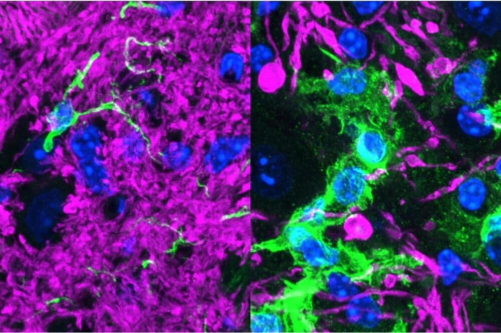
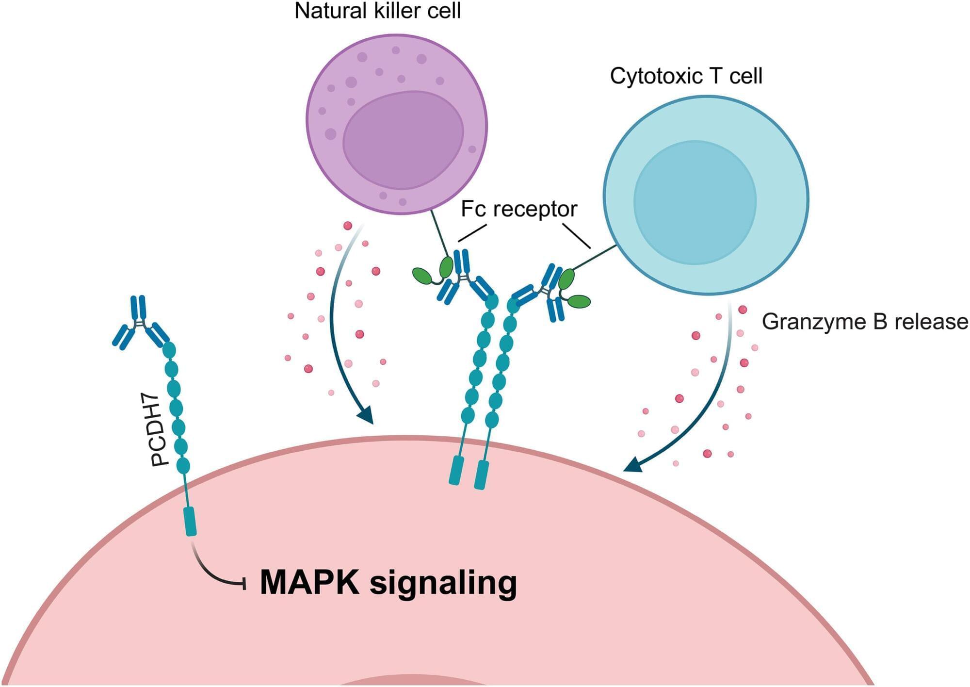
An experimental antibody treatment that binds to a protein known as PCDH7 shrank tumors in preclinical models of non-small cell lung cancer (NSCLC), including those resistant to a targeted therapy, a study led by UT Southwestern Medical Center researchers showed. The findings, published in Science Advances, could eventually lead to a new class of drugs to treat NSCLC and potentially other cancers.

“Overcoming resistance to molecularly targeted therapies is a critical unmet need for lung cancer patients. We are excited that these antibodies may open another therapeutic avenue for lung cancer, especially for patients whose cancers have become resistant to KRAS inhibitors,” said Kathryn O’Donnell, Ph.D., associate professor of molecular biology and a member of the Harold C. Simmons Comprehensive Cancer Center at UT Southwestern. O’Donnell co-led the study with first author Nicole Novaresi, Ph.D., a postdoctoral researcher in the O’Donnell Lab, and collaborators at the University of Texas Health Science Center at Houston.

NSCLC accounts for about 85% of lung cancer cases in the U.S. and is the leading cause of cancer-related deaths. The O’Donnell Lab focuses on identifying and characterizing proteins on the surface of NSCLC and other cancer cells because of their potential as therapeutic targets. In 2017, O’Donnell and her colleagues identified PCDH7 as a driver of NSCLC, especially in tumors with mutations in a gene called KRAS. Found in about 25% of NSCLC cases, these mutations cause uncontrolled cell proliferation that propels tumor growth.
How the body remembers the tumor?

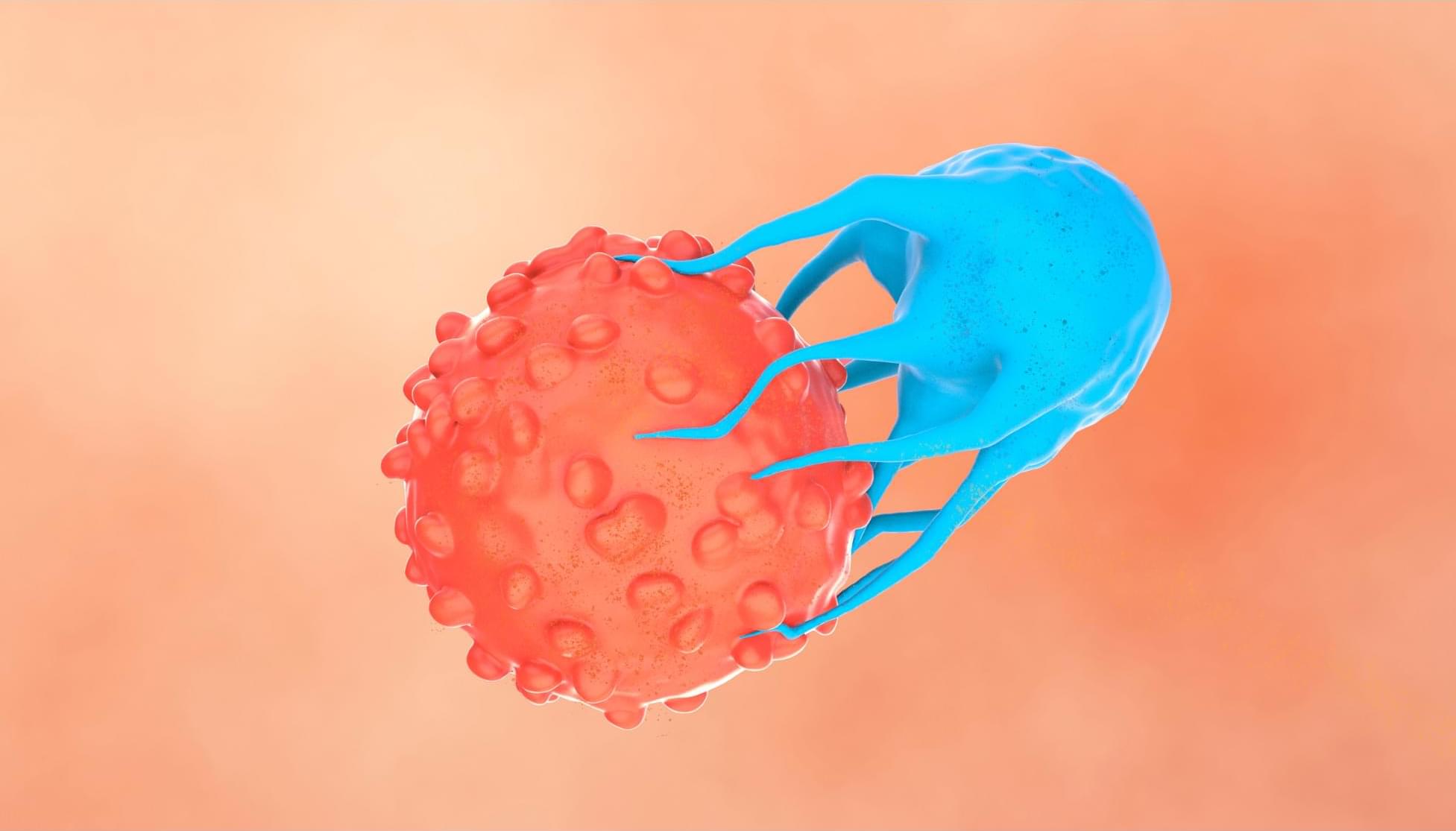
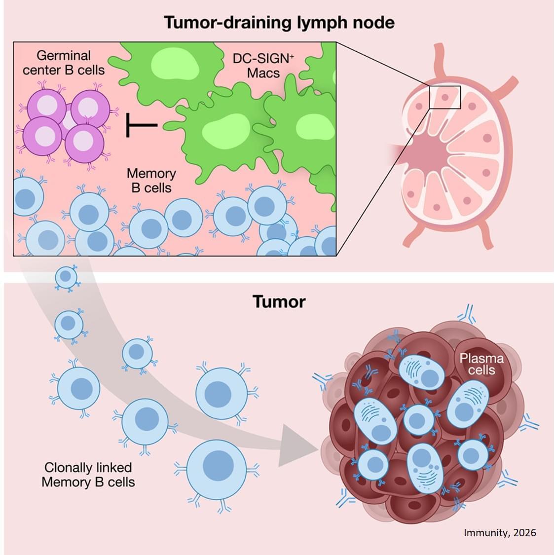
While we tend to quickly forget having been ill or having received a vaccine, the immune system remembers remarkably well. It has memory B cells – “trained” immune cells that circulate throughout the body in search of harmful invaders they have encountered previously; these cells can rapidly deploy targeted weapons when faced with a pathogen again. Now, researchers report that activated memory B cells can also recognize an internal enemy: cancer cells.

In patients with ovarian cancer, the researchers identified memory cells that are capable of homing in on the tumor, springing into action and producing effective antibodies against it. The new study, whose findings were published in Immunity, advances the development of vaccines and therapies based on immune memory against cancer.

The immune system’s arsenal contains hundreds of millions of B cell clones, each producing a unique antibody against a specific pathogen. These antibodies are proteins that identify their target and either neutralize it or recruit other immune cells to attack it. When a clone encounters its target for the first time, its antibody binds weakly and elicits a limited response. But some of these cells enter “training camps” – structures called germinal centers in the lymph nodes – where they undergo genetic changes and rigorous selection, emerging with much more effective antibodies. Some of these trained cells immediately become active antibody producers; others develop into memory cells that remain inactive, circulating between the blood and the lymph nodes, but able to rapidly snap into action if the body is exposed again to the pathogen.

Faster biological aging consistently linked to poverty and discrimination

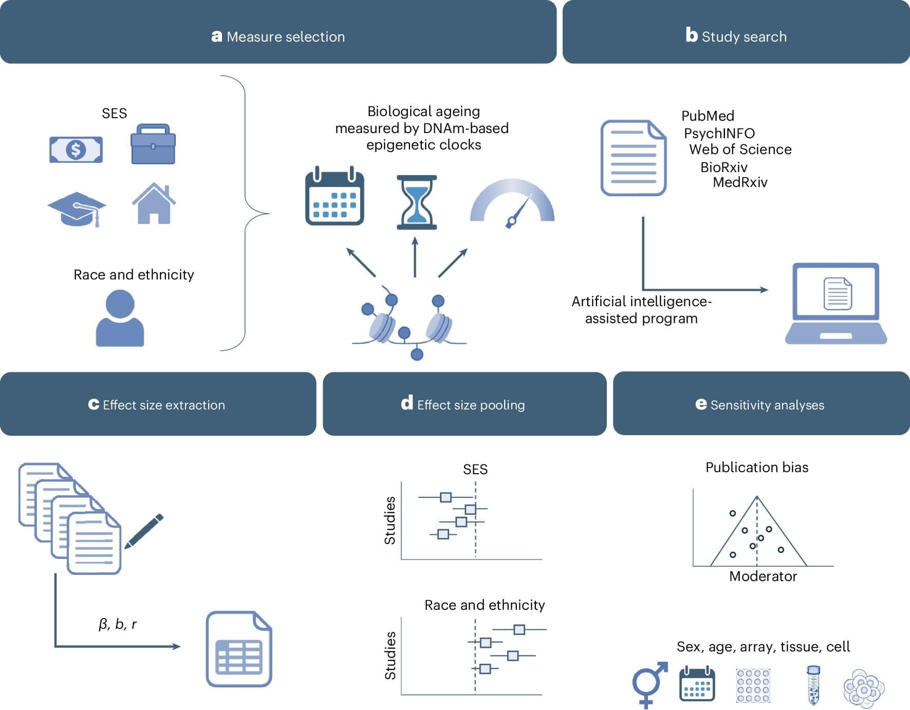
The study, published in Nature Human Behaviour, demonstrates that social inequality, such as poverty and racism, is related to biological aging measured in the epigenome, also known as “epigenetic clocks.” Epigenetic clocks analyze patterns of chemical marks on DNA to estimate a person’s biological age or the rate at which their body is aging. These tools are increasingly used by scientists to study how environmental exposures, lifestyle and social conditions affect health across the life course.

Previous individual studies have shown that epigenetic clocks are sensitive to socioeconomic and racial or ethnic disparities. However, because multiple types of epigenetic clocks exist, it has remained unclear which measures best capture the effects of social determinants of health, at which stages of life socioeconomic exposures most affect epigenetic aging, and whether associations differ by sex or by technical factors such as the tissue in which epigenetic data are collected. This study integrates findings across many independent studies, offering a comprehensive test of whether these associations are consistent and robust.

Faulty protein cleanup gene tied to severe early-onset neurological disorders

Though protein clumps associated with Alzheimer’s and Parkinson’s were discovered more than a century ago, researchers remain largely unable to prevent them from forming or eliminate them from the brain. And though a variety of therapies have taken aim at tau tangles, beta-amyloid plaques and Lewy bodies, among other notorious aggregates, none have been very effective at stopping disease progression.

Rockefeller’s Hermann Steller and his team in the Strang Laboratory of Apoptosis and Cancer Biology have long been focused on understanding how the cell’s protein-degrading machines, called proteasomes, are regulated. His lab discovered that a transporter protein termed PI31 shuttles proteasomes over long distances from the nerve cell body to synapses. When this system fails, synapses become depleted of degradative capacity, and proteins that should have been eliminated accumulate. As a result, synaptic communication breaks down, protein clumps form and neuronal health deteriorates.

Now a new study in Nature Communications, led by researchers from University College London and contributed to by Steller’s lab, has identified mutations in PSMF1, the gene that produces PI31, that cause the protein to malfunction. Moreover, the scientists demonstrated that these mutations cause a spectrum of severe, very early-onset neurological disorders.

Making CAR T Cells Safer

Research from CCR scientists points toward a strategy for making chimeric antigen receptor (CAR) T-cell therapy, the cell-based immunotherapy that has revolutionized the treatment of some blood cancers, safer and more effective for treating solid tumors.

The study, led by Grégoire Altan-Bonnet, Ph.D., Deputy Chief of the Laboratory of Integrative Cancer Immunology, Naomi Taylor, M.D., Ph.D., Senior Investigator in the Pediatric Oncology Branch, and Paul François, Ph.D., at the University of Montréal, shows how adding certain receptors to CAR T cells can prevent the cells from attacking healthy tissue while simultaneously enhancing their activity against cancer cells. The findings appeared April 10, 2025, in Cell.

CAR T-cell therapy reprograms patients’ immune cells to be effective cancer killers using genetically engineered chimeric antigen receptors (CARs) that are added to their T cells. CARs are designed to recognize molecules on the surface of cancer cells called antigens, which can usually be found on some healthy cells, too. This leads to manageable side effects for patients with blood cancer, but when CAR T cells designed to target solid tumors attack healthy tissue, the effects can be severe.

Genomes from Oceania offer new clues to human evolution

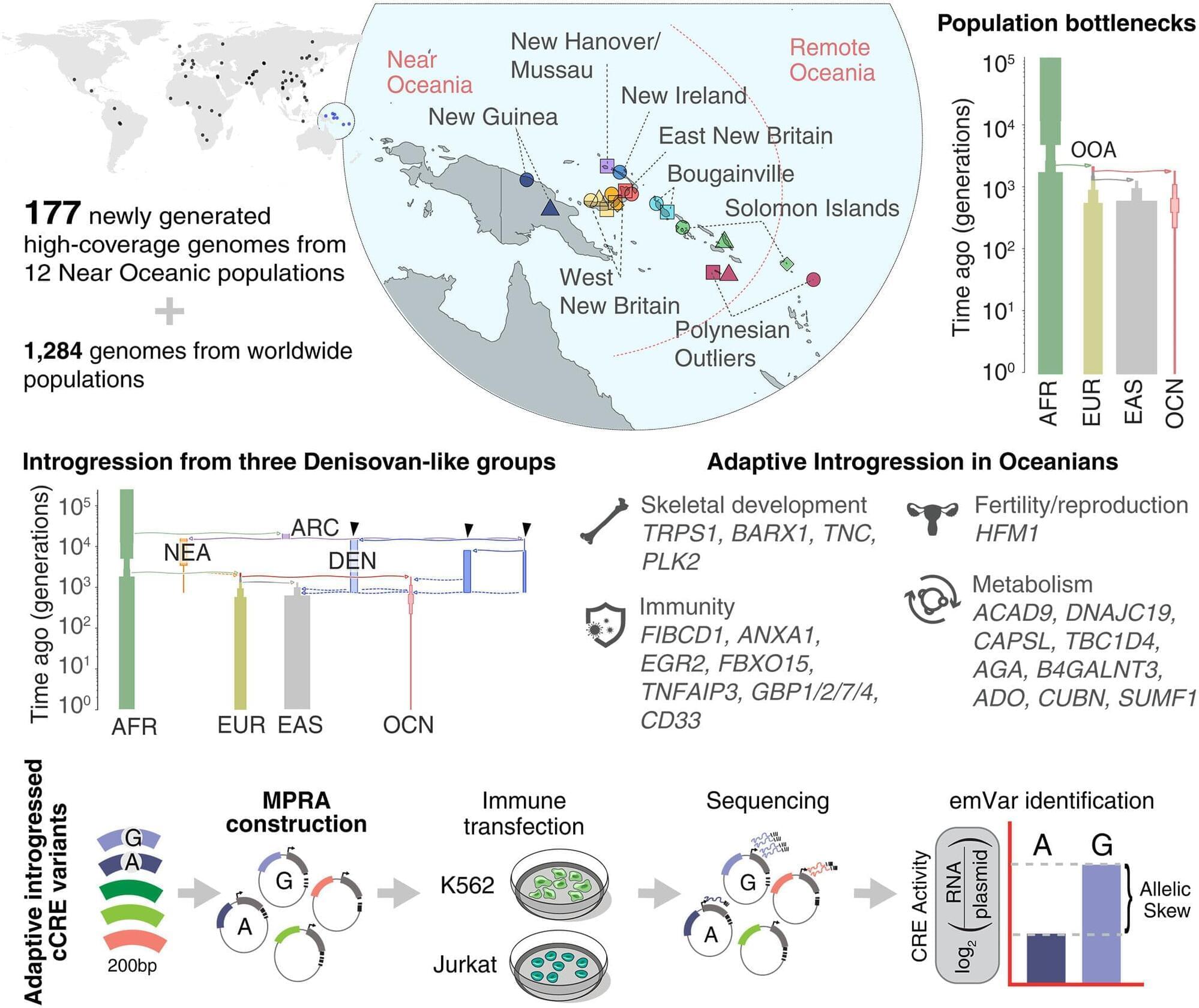
A new Yale-led study provides one of the most detailed and comprehensive analyses to date of genetic variation in human populations in Oceania, filling a major gap in representation in genomics research. Despite harboring remarkable diversity, populations in this vast region in the South Pacific historically have been overlooked in global human genetic studies, which have often focused largely on people of European descent, researchers say. The study is published in the journal Science.

“The drastic underrepresentation of Oceanians limits our understanding of human evolution and could exacerbate health inequalities as genomic research is used to develop novel medical treatments,” said lead author Serena Tucci, assistant professor of anthropology in Yale’s Faculty of Arts and Sciences and principal investigator of the Yale Human Evolutionary Genomics Laboratory. “To fill that gap, my research team embarked on a large-scale project to expand what is known about human genetic variation, including genetic variants inherited from extinct hominins.”

The work shows how the genes that ancient humans acquired after mating with extinct hominins continue to shape the biology, health and survival of our species today.

CRISPR enzyme precisely detects and shreds DNA in cancer mutations once considered ‘undruggable’

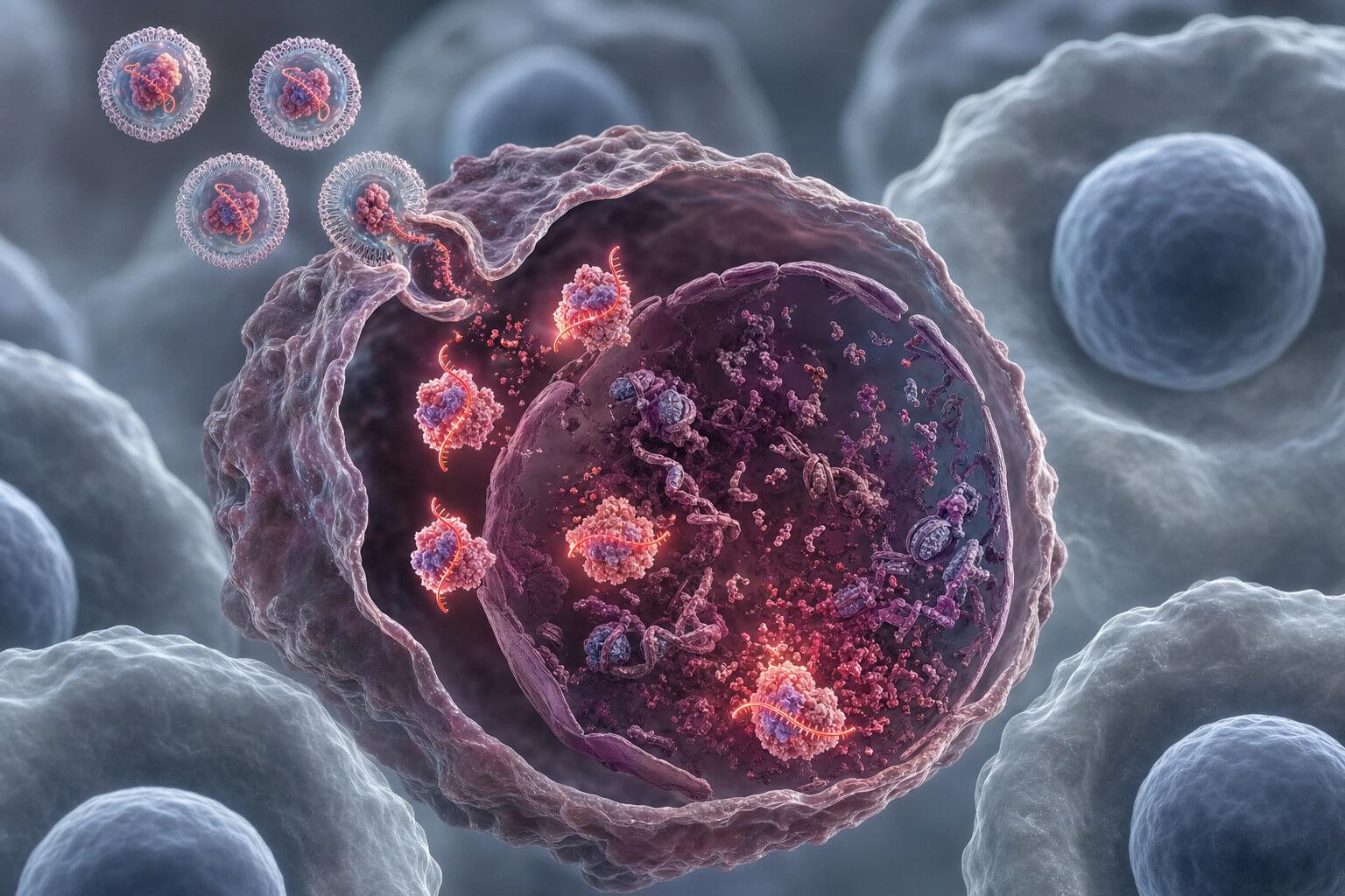
In 2020, Jennifer Doudna won the Nobel Prize in chemistry for her work on the CRISPR-Cas9 gene-editing technology that allows scientists to precisely modify DNA by cutting it at specific locations. Six years later, a new study in Nature by a team led by Doudna has uncovered a powerful new approach to selectively kill cancer cells using a CRISPR enzyme called Cas12a2.

Once the enzyme detects cancer-specific genetic signatures, it begins to shred chromatin—a mixture of DNA and proteins that forms chromosomes—within the targeted cell.

Many cancers are driven by mutations in tumor suppressor proteins such as TP53, which is altered in nearly half of all cases. Yet these mutations have remained difficult to treat because they lack binding pockets for traditional drugs to latch onto. As a result, many cancer-causing mutations have long been considered undruggable.

Silver nanoparticles pave the way for precise DNA cutting and joining

DNA is composed of long chains that act as the blueprint for living organisms. In genetic engineering, scientists cut DNA at specific sites and join the resulting fragments to other DNA sequences, enabling applications such as advanced crop breeding, treatment of genetic diseases, and the generation of animal models for drug discovery.

Assembling short DNA fragments requires overhanging sequences, known as sticky ends, to facilitate efficient binding. However, generating sticky ends requires precise cutting at targeted sites, which remains challenging with current technologies.

A Japanese research group has developed a silver nanoparticle-based technology to precisely cut and join DNA at targeted sites, achieving two to five times higher DNA assembly efficiency than conventional restriction enzyme methods. These findings were published in the journal Nucleic Acids Research.

Researchers trigger sleep’s restorative effect in parts of the awake brain

Scientists from the University of Wisconsin-Madison have successfully replicated some of the restorative effects of deep sleep in awake mice by artificially inducing slow-wave brain activity. Using optogenetics to control specific neurons, researchers triggered localized cortical activity that mimics the NREM sleep phase responsible for synaptic homeostasis and the reorganization of neural connections. This targeted stimulation significantly reduced signs of fatigue and improved memory retention and cognitive performance in the mice following prolonged wakefulness. While the researchers caution that this technique is not a substitute for natural sleep, the findings suggest that localized neural stimulation can effectively preserve brain function during extended periods of wakefulness. Future research aims to explore whether similar cognitive benefits can be achieved in humans through non-invasive methods, such as transcranial electrical stimulation.


NIH-funded study in animals offers new details about how the brain resets during sleep.

By inducing specific patterns of activity in small portions of the brain in awake mice, researchers supported by the National Institutes of Health (NIH) have triggered a recalibration of neural connections that normally only occurs during sleep. This new approach offset the effects of sleep deprivation in memory tasks and revealed features of sleep that are key to its restorative effect.

“What we’re essentially doing is forcing sleep in a local region of the brain. While that part is solidifying memories and restoring learning capacity, other parts stay aware/vigilant and connected to environment,” said corresponding author Chiara Cirelli, M.D., Ph.D., a professor of psychiatry at the University of Wisconsin-Madison. “Dolphins do something similar, sleeping with only one brain hemisphere at a time.”

/* */