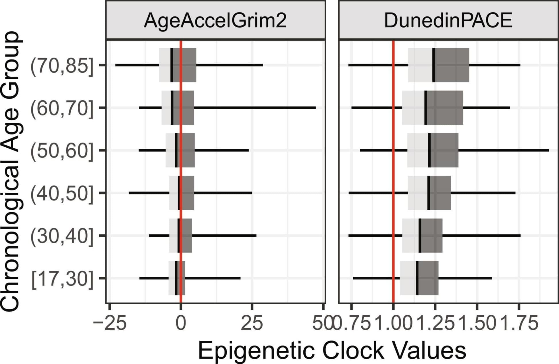
Toggle light / dark theme

Northwestern Medicine investigators have discovered new molecular mechanisms underlying DNA repair dysregulation in prostate cancer cells, findings that may inform the development of new targeted therapies for patients that have become resistant to standard treatments, according to a recent study published in Science Advances.

Qi Cao, Ph.D., the Anthony J. Schaeffer, MD, Professor of Urology, was senior author of the study.

DNA damage is a natural occurrence in cells caused by various intercellular and external stressors. However, if left unrepaired, this damage can lead to genetic mutations that can lead to the development of different diseases, including cancer.

Within a cell, DNA carries the genetic code for building proteins. To build proteins, the cell makes a copy of DNA, called mRNA. Then, another molecule called a ribosome reads the mRNA, translating it into protein. But this step has been a visual mystery; scientists previously did not know how the ribosome attaches to and reads mRNA.

Now, a team of international scientists, including University of Michigan researchers, has used advanced microscopy to image how ribosomes recruit to mRNA while it’s being transcribed by an enzyme called RNA polymerase (RNAP). Their results, which examine the process in bacteria, are published in the journal Science.

“Understanding how the captures or ‘recruits’ the mRNA is a prerequisite for everything that comes after, such as understanding how it can even begin to interpret the information encoded in the mRNA,” said Albert Weixlbaumer, a researcher from Institut de génétique et de biologie moléculaire et cellulaire in France who co-led the study.

Resistance to one antibiotic can make bacteria resistant or sensitive to another antibiotic, opening paths for combinatorial treatments. This study presents an approach to systematically discover and understand such antibiotic relationships.

Join us on Patreon! https://www.patreon.com/MichaelLustgartenPhD

Discount Links/Affiliates:
Blood testing (where I get the majority of my labs): https://www.ultalabtests.com/partners/michaellustgarten.

At-Home Metabolomics: https://www.iollo.com?ref=michael-lustgarten.
Use Code: CONQUERAGING At Checkout.

Clearly Filtered Water Filter: https://get.aspr.app/SHoPY

Scientists from the Longevity Research Institute (LRI), which was formed by the merger of SENS Research Foundation and Lifespan.io, have achieved expression of an essential mitochondrial gene in the nucleus and proper functioning of the protein. This could pave the way for curing diseases caused by mitochondrial mutations [1].

The fragile mitochondrial DNA

The prevailing scientific consensus is that mitochondria were once independent microorganisms that entered a symbiotic relationship with larger cells. This duo gave rise to eukaryotic cells: the building blocks of all multicellular life. Without that fateful “marriage,” complex life would not exist, as mitochondria provide cells with essential energy via oxidative phosphorylation.

Cleveland Clinic Genome Center researchers have unraveled how immune cells called microglia can transform and drive harmful processes like neuroinflammation in Alzheimer’s disease. The study, published in the journal Alzheimer’s & Dementia, also integrates drug databases with real-world patient data to identify FDA-approved drugs that may be repurposed to target disease-associated microglia in Alzheimer’s disease without affecting the healthy type.

The researchers, led by study corresponding author Feixiong Cheng, Ph.D., hope their unique approach of integrating genetic, chemical and human health data to identify and corresponding drugs will inspire other scientists to take similar approaches in their own research.

Microglia are specialized that patrol our brains, seeking and responding to tissue damage and external threats like bacteria and viruses. Different types of microglial cells use different methods to keep the brain safe. Some may cause neuroinflammation—inflammation in the brain—to fight invaders or kickstart the repair process in damaged cells. Others may work to “eat” dangerous substances in the brain, and clean up damage and debris. However, during Alzheimer’s disease, new types of microglia can form that promote .

Its a problem, but im sure ASI by 2035 will solve for a way to use a Crispr type tool with zero unintended alterations. Look for a way to use w/ out alterations in meantime, but worst case ASI will solve it.


Genome editing with various CRISPR-Cas molecule complexes has progressed rapidly in recent years. Hundreds of labs around the world are now working to put these tools to clinical use and are continuously advancing them.

CRISPR-Cas tools allow researchers to modify individual building blocks of genetic material in a precise and targeted manner. Gene therapies based on such gene editing are already being used to treat inherited diseases, fight cancer and create drought-and heat-tolerant crops.

The CRISPR-Cas9 molecular complex, also known as genetic scissors, is the most widely used tool by scientists around the world. It cuts the double-stranded DNA at the exact site where the genetic material needs to be modified. This contrasts with newer gene-editing methods, which do not cut the double strand.

A recent study investigates the relationship between exercise and the expression of MYC in skeletal muscles over time, revealing that even minimal doses can promote muscle growth without physical activity.

Researchers have long known that there is a relationship between the cancer-associated gene MYC (pronounced “Mick”) and exercise adaptation. When human muscles are exercised, MYC is found to increase transiently in abundance over 24 hours. But as we age, the MYC response to exercise is blunted, perhaps explaining a reduced ability to recover from exercise and maintain or gain muscle.

Knowing the precise mechanisms by which MYC drives muscle growth could prove instrumental in creating therapies that reduce muscle loss from aging, potentially improving independence, mobility, and health.