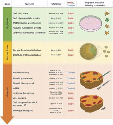
Toggle light / dark theme

New research has uncovered how genetic changes that accumulate slowly in blood stem cells throughout life are likely to be responsible for the dramatic change in blood production after the age of 70.

The study, by scientists at the Wellcome Sanger Institute, the Wellcome-MRC Cambridge Stem Cell Institute and collaborators, has been published in the journal Nature.

Longevity. Technology: Has our understanding of one of the mechanisms of aging taken a quantum leap? Molecular damage accumulates throughout our lives, gradually increasing year-on-year as we suffer telomere attrition, mutation, epigenetic change and oxidative and replicative stress. It’s a double whammy as our ability to repair this damage also declines as we age, but given the gradual nature of these processes, why, as the paper authors themselves put it, “Is there an abrupt increase in mortality after 70 years of age? [1].

“Functional mutations in the growth hormone pathway” meaning it is not active. What’s good for you as a youngster might not be good for you when you’re old.


Dr Nir Barzilai reveals what the longevity genes project found on why Centenarians live longer, not the longevity genes, not healthy lifestyles in this clip.

Dr. Nir Barzilai is the director of the Institute for Aging Research at the Albert Einstein College of Medicine and the Director of the Paul F. Glenn Center for the Biology of Human Aging Research and of the National Institutes of Health’s (NIH) Nathan Shock Centers of Excellence in the Basic Biology of Aging. He is the Ingeborg and Ira Leon Rennert Chair of Aging Research, professor in the Departments of Medicine and Genetics, and member of the Diabetes Research Center and of the Divisions of Endocrinology & Diabetes and Geriatrics.

A drug combination targeting multiple mutant versions of cancer’s “death star” protein has shown promise in a small, early-phase clinical trial for some patients with advanced lung, ovarian and thyroid cancer.

The two– was effective against with a range of mutations to the KRAS gene—dubbed the “death star” because its protein drives one in four cancers and has a largely impenetrable, drug-resistant surface.

The phase I trial tested the drugs VS-6766 and everolimus in 30 patients with a range of mutations to KRAS—including 11 with highly advanced, .

Observer, backup youthful copy, playing the right piano notes, quantum states oh my.


Dr David Sinclair explain about through his lab experiments, why he thinks there is an observer/backup copy for our youthfulness and what are the possible identities he can think of in this clip.

David Sinclair is a professor in the Department of Genetics and co-director of the Paul F. Glenn Center for the Biology of Aging at Harvard Medical School, where he and his colleagues study sirtuins—protein-modifying enzymes that respond to changing NAD+ levels and to caloric restriction—as well as chromatin, energy metabolism, mitochondria, learning and memory, neurodegeneration, cancer, and cellular reprogramming.

Scientists at the Foundation for Applied Molecular Evolution announced today that ribonucleic acid (RNA), an analog of DNA that was likely the first genetic material for life, spontaneously forms on basalt lava glass. Such glass was abundant on Earth 4.35 billion years ago. Similar basalts of this antiquity survive on Mars today.


More information:

Craig A. Jerome et al, Catalytic Synthesis of Polyribonucleic Acid on Prebiotic Rock Glasses, Astrobiology (2022). DOI: 10.1089/ast.2022.

Hyo-Joong Kim et al, Prebiotic stereoselective synthesis of purine and noncanonical pyrimidine nucleotide from nucleobases and phosphorylated carbohydrates, Proceedings of the National Academy of Sciences (2017). DOI: 10.1073/pnas.

Scientists have produced the first genetic map of chimpanzees in the wild, offering a detailed reconstruction of the endangered species’ past migrations, and a new tool to combat illegal trafficking.

The genomic catalogue, which includes 828 individuals from across their vast African range, can now be used to link kidnapped chimpanzees—or their meat and —to their place of origin within 100 kilometers.

The results of the years-long research project was published Wednesday in the journal Cell Genomics.

Circa 2019 immortality in the human brain 🧠


Brain injuries causing chronic sensory or motor deficit, such as stroke, are among the leading causes of disability worldwide, according to the World Health Organization; furthermore, they carry heavy social and economic burdens due to decreased quality of life and need of assistance. Given the limited effectiveness of rehabilitation, novel therapeutic strategies are required to enhance functional recovery. Since cell-based approaches have emerged as an intriguing and promising strategy to promote brain repair, many efforts have been made to study the functional integration of neurons derived from pluripotent stem cells (PSCs), or fetal neurons, after grafting into the damaged host tissue. PSCs hold great promises for their clinical applications, such as cellular replacement of damaged neural tissues with autologous neurons. They also offer the possibility to create in vitro models to assess the efficacy of drugs and therapies. Notwithstanding these potential applications, PSC-derived transplanted neurons have to match the precise sub-type, positional and functional identity of the lesioned neural tissue. Thus, the requirement of highly specific and efficient differentiation protocols of PSCs in neurons with appropriate neural identity constitutes the main challenge limiting the clinical use of stem cells in the near future. In this Review, we discuss the recent advances in the derivation of telencephalic (cortical and hippocampal) neurons from PSCs, assessing specificity and efficiency of the differentiation protocols, with particular emphasis on the genetic and molecular characterization of PSC-derived neurons. Second, we address the remaining challenges for cellular replacement therapies in cortical brain injuries, focusing on electrophysiological properties, functional integration and therapeutic effects of the transplanted neurons.

Brain injuries represent a large variety of disabling pathologies. They may originate from different causes and affect distinct brain locations, leading to an enormous multiplicity of various symptoms ranging from cognitive deficits to sensorimotor disabilities. They can also result in secondary disturbances, such as epileptic foci, which occur within the lesioned and perilesional tissues (Herman, 2002). Indeed, frequently a secondary functional damage can take place in a region distant from the first insult (e.g., the hippocampus after traumatic brain injury), providing an explanation for cognitive and memory deficits arising after a brain lesion (Girgis et al., 2016). Brain injuries can have traumatic or non-traumatic etiologies, including focal brain lesions, anoxia, tumors, aneurysms, vascular malformations, encephalitis, meningitis and stroke (Teasell et al., 2007). In particular, stroke covers a vast majority of acquired brain lesions.