1Health.io left its customers genetic information unsecured in unencrypted and publicly accessible AWS servers.

Human lifespan is shaped by both genetic and environmental exposures and their interaction. To enable precision health, it is essential to understand how genetic variants contribute to earlier death or prolonged survival. In this study, we tested the association of common genetic variants and the burden of rare non-synonymous variants in a survival analysis, using age-at-death (N = 35,551, median [min, max] = 72.4 [40.9, 85.2]), and last-known-age (N = 358,282, median [min, max] = 71.9 [52.6, 88.7]), in European ancestry participants of the UK Biobank. The associations we identified seemed predominantly driven by cancer, likely due to the age range of the cohort. Common variant analysis highlighted three longevity-associated loci: APOE, ZSCAN23, and MUC5B. We identified six genes whose burden of loss-of-function variants is significantly associated with reduced lifespan: TET2, ATM, BRCA2, CKMT1B, BRCA1 and ASXL1. Additionally, in eight genes, the burden of pathogenic missense variants was associated with reduced lifespan: DNMT3A, SF3B1, CHL1, TET2, PTEN, SOX21, TP53 and SRSF2. Most of these genes have previously been linked to oncogenic-related pathways and some are linked to and are known to harbor somatic variants that predispose to clonal hematopoiesis. A direction-agnostic (SKAT-O) approach additionally identified significant associations with C1orf52, TERT, IDH2, and RLIM, highlighting a link between telomerase function and longevity as well as identifying additional oncogenic genes.
Our results emphasize the importance of understanding genetic factors driving the most prevalent causes of mortality at a population level, highlighting the potential of early genetic testing to identify germline and somatic variants increasing one’s susceptibility to cancer and/or early death.
The authors have declared no competing interest.
Sources and further reading:
- Bridge RNAs direct programmable recombination of target and donor DNA https://www.nature.com/articles/s4158…
- Structural mechanism of bridge RNA-guided recombination https://www.nature.com/articles/s4158…
Join us on Patreon! https://www.patreon.com/MichaelLustgartenPhDDiscount Links/Affiliates (Ways To Test Yourself, While Also Helping To Support The Channe):…
This gene therapy treats LCA1, causing early childhood vision loss, affecting under 100,000 people:
“One patient reported for the first time being able to navigate at midnight outdoors only with the light of a bonfire,” said Cideciyan, who is also co-director of the Center for Hereditary Retinal Degenerations.
The clinical trials were co-led by researchers from the Perelman School of Medicine at the University of Pennsylvania.
The gene therapy (ATSN-101) is specifically designed to target and correct the genetic mutation in the GUCY2D gene. This gene creates vision-imparting proteins. ATSN-101 is “adapted from the AAV5 microorganism.”


Summary: A new gene therapy has restored vision in patients with Leber congenital amaurosis type I (LCA1), a rare genetic condition causing blindness. In a small trial, those receiving the highest dose saw up to a 10,000-fold improvement in light sensitivity and significant gains in reading and navigation abilities.
The therapy, developed by researchers, uses a virus-based system to deliver a functioning gene into the retina’s light-sensitive cells. The results show promise for expanding this treatment, with further trials planned to confirm safety and efficacy.

The vision of people with a rare inherited condition that causes them to lose much of their sight early in childhood was 100 times better after they received gene therapy to address the genetic mutation causing it. Some patients even experienced a 10,000-fold improvement in their vision after receiving the highest dose of the therapy, according to researchers from the Perelman School of Medicine at the University of Pennsylvania who co-led the clinical trial published in The Lancet.
“That 10,000-fold improvement is the same as a patient being able to see their surroundings on a moonlit night outdoors as opposed to requiring bright indoor lighting before treatment,” said the study’s lead author, Artur Cideciyan, Ph.D., a research professor of Ophthalmology and co-director of the Center for Hereditary Retinal Degenerations.
“One patient reported for the first time being able to navigate at midnight outdoors only with the light of a bonfire.”
Their…
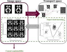
A multi-university research team co-led by University of Virginia engineering professor Gustavo K. Rohde has developed a system that can spot genetic markers of autism in brain images with 89 to 95% accuracy.
Their findings suggest doctors may one day see, classify and treat autism and related neurological conditions with this method, without having to rely on, or wait for, behavioral cues. And that means this truly personalized medicine could result in earlier interventions.
“Autism is traditionally diagnosed behaviorally but has a strong genetic basis. A genetics-first approach could transform understanding and treatment of autism,” the researchers wrote in a paper published June 12 in the journal Science Advances.