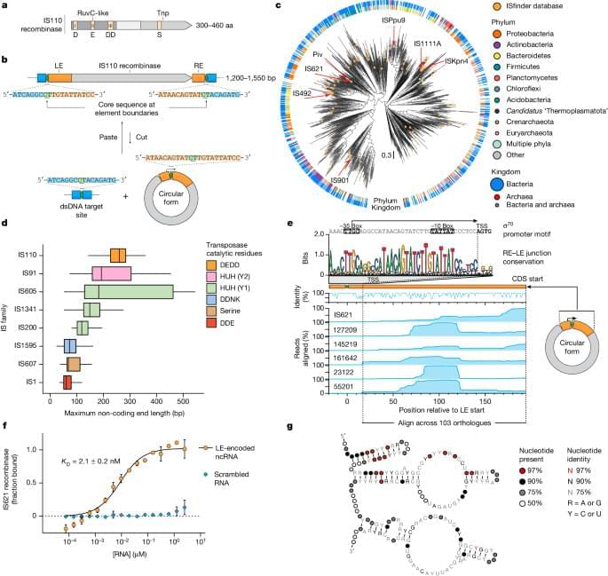
Check out this astonishing publication by Durrant et al.
A bispecific non-coding RNA expressed by the IS110 family of mobile genetic elements forms the basis of a programmable genome-editing system that enables the insertion, excision or inversion of specific target DNA sequences.
The Road To Wisdom — Dr. Francis Collins, MD, PhD — Former Director, National Institutes of Health (NIH); Distinguished Investigator, Center for Precision Health Research, National Human Genome Research Institute.
Dr. Francis S. Collins, M.D., Ph.D., (https://www.francisscollins.com/) is the former Director of the U.S. National Institutes of Health (NIH), where as the longest serving director of NIH (spanning 12 years and three presidencies) he oversaw the work of the largest supporter of biomedical research in the world, from basic to clinical research.
Dr. Collins continues to serve as NIH Distinguished Investigator. Center for Precision Health Research, at the National Human Genome Research Institute (NHGRI — https://irp.nih.gov/pi/francis-collins).
Dr. Collins is a physician-geneticist noted for his landmark discoveries of disease genes and his leadership of the international Human Genome Project, which culminated in April 2003 with the completion of a finished sequence of the human DNA instruction book. He served as director of the National Human Genome Research Institute at the NIH from 1993–2008.
Dr. Collins’ research laboratory has discovered a number of important genes, including those responsible for cystic fibrosis, neurofibromatosis, Huntington’s disease, a familial endocrine cancer syndrome, and most recently, genes for type 2 diabetes, and the gene that causes Hutchinson-Gilford progeria syndrome, a rare condition that causes premature aging.
In a new study, scientists from Arizona State University and their collaborators studied genetic changes in a naturally isolated population of Daphnia pulex, a species of water flea. This tiny crustacean, nearly invisible to the naked eye, plays a vital role in freshwater ecosystems and provides a valuable insight into natural selection and evolution.
Their findings, recently published in the journal Proceedings of the National Academy of Sciences (PNAS), rely on a decade of research. Using advanced genomic techniques, the research team analyzed DNA samples from nearly 1,000 Daphnia.
They discovered that the strength of natural selection on individual genes varies significantly from year to year, maintaining variation and potentially enhancing the ability to adapt to future changing environmental conditions by providing raw material for natural selection to act on.
Irrespective of their personal, professional and social circumstances, different individuals can experience varying levels of life satisfaction, fulfillment and happiness. This general measure of life satisfaction, broadly referred to as “well-being,” has been the key focus of numerous psychological studies.
Better understanding the many factors contributing to well-being could help to devise personalized and targeted interventions aimed at improving people’s levels of fulfillment. While many past studies have tried to delineate these factors, few have done so leveraging the advanced machine learning models available today.
Machine learning models are designed to analyze large amounts of data, unveiling hidden patterns and making accurate predictions. Using these tools to analyze data collected in previous studies in neuroscience and psychology could help to shed light on the environmental and genetic factors influencing well-being.
Marfan syndrome is a genetic disorder that leads to a greater risk of aneurysms developing in a patient’s aorta. Early detection is key to survival. Researchers at Yale School of Medicine studied the use of artificial intelligence in the diagnostic process. The results could eventually lead to an easier and more accessible test.
Have we created an operating system for life? How close are we to cloning humans, and what would that even look like?
You’re in for a fascinating episode as the line between science and science fiction gets blurred. My guest is microbiologist and geneticist Andrew Hessel, the CEO and Founder of The Genome Project-Write, and author of \.
Life insurers and those offering income protection and permanent disability insurance will be banned from using genetic testing to refuse cover, or hike up charges, for a range of insurance products.
The federal government announced on Tuesday it would ban the practice that saw consumers discriminated against if they disclosed the results of genetic tests that predict their likelihood of an inherited disease.
It comes after consultation to address genetic discrimination in life insurance earlier this year. More than 1,000 submissions were received with 97 per cent supporting a total ban.
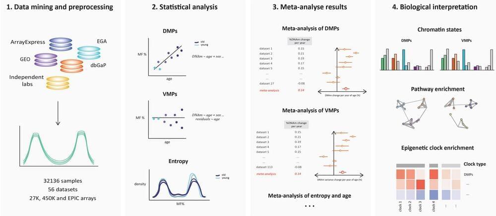
During aging, the human methylome undergoes both differential and variable shifts, accompanied by increased entropy. The distinction between variably methylated positions (VMPs) and differentially methylated positions (DMPs), their contribution to epigenetic age, and the role of cell type heterogeneity remain unclear.
We conduct a comprehensive analysis of 32,000 human blood methylomes from 56 datasets (age range = 6–101 years). We find a significant proportion of the blood methylome that is differentially methylated with age (48% DMPs; FDR 0.005) and variably methylated with age (37% VMPs; FDR 0.005), with considerable overlap between the two groups (59% of DMPs are VMPs). Bivalent and Polycomb regions become increasingly methylated and divergent between individuals, while quiescent regions lose methylation more uniformly. Both chronological and biological clocks, but not pace-of-aging clocks, show a strong enrichment for CpGs undergoing both mean and variance changes during aging. The accumulation of DMPs shifting towards a methylation fraction of 50% drives the increase in entropy, smoothening the epigenetic landscape. However, approximately a quarter of DMPs exhibit anti-entropic effects, opposing this direction of change.