Toggle light / dark theme

Naked mole rats are rodents that are about the size of a mouse with a key difference, aside from having no fur — they’re extremely long-lived — reaching ages of around 40 years old. For comparison, lab mice live an average of about three and a half years. To explain their extensive lifespans, researchers have sought to pinpoint how naked mole rats evade the onset of age-related diseases like cancer. In doing so, they’ve identified a form of gelatinous substance called hyaluronan, which has anti-inflammatory and anticancer properties. Now, the question of whether the benefits of the naked mole rat’s abundant levels of this form of hyaluronan — called high molecular mass hyaluronic acid (HMM-HA) — can be exported to other species has recently drawn attention.

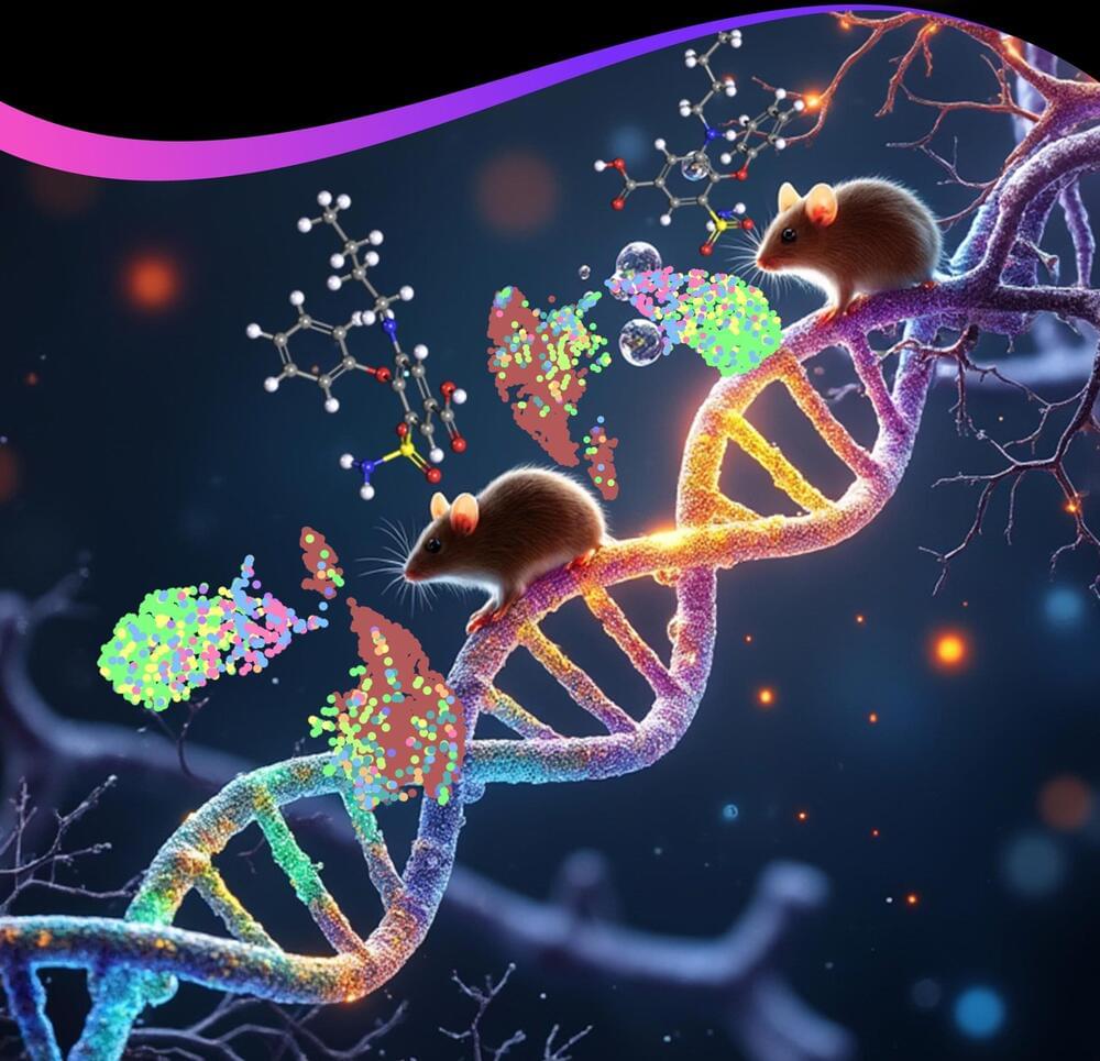
Published in Nature, Gorbunova and colleagues from the University of Rochester show that genetically modifying mice to harbor an enzyme that produces HMM-HA extends their lifespan. The researchers go on to show that increasing HMM-HA reduces the prevalence of cancer. Additionally, the nmrHAS2 gene improves the healthspan of mice by countering physiological dysfunction, as measured with a frailty score. These findings provide the first evidence that genes from long-lived species can be exported to other species, perhaps conferring benefits to humans one day.

Plants possess the unique ability to completely regenerate from a somatic cell, i.e., an ordinary cell that does not typically participate in reproduction. This process involves the de novo (or new) formation of a shoot apical meristem (SAM) that gives rise to lateral organs, which are key for the plant’s reconstruction.

On a cellular scale, the formation of SAM is meticulously controlled by either positive or negative regulators (genes/protein molecules) that may induce or restrict shoot regeneration, respectively. But which molecules are involved? Are there other regulatory layers that are yet to be uncovered?

To seek answers to the above questions, a research group led by Nara Institute of Science and Technology (NAIST), Japan studied the process in Arabidopsis, a plant commonly used in genetic research.

In this article, the fourth installment of our five-part series on different pathways of aging, we look at the rejuvenation of cells, tissues, and stem cells, a topic that has been gaining increasing popularity thanks to remarkable advancements in the field of epigenetic reprogramming. Recent research suggests that despite the accumulation of molecular damage over time, cells and tissues can indeed undergo rejuvenation. We’ll be exploring key subjects such as Epigenetic reprogramming, PGC1a and GSK3β, Telomerase (TERT), as well as Apoptosis and senescence. Join us on this enlightening journey as we uncover the groundbreaking discoveries that are shaping the future of aging research.

The idea for reprogramming was simple yet beautiful. Children are born young, even though their parents are old, because they have undergone a process of cellular reprogramming that leads to rejuvenation.

In a recent study published in the Journal of Experimental Medicine, researchers investigated whether bone marrow-derived cells with heterozygous loss of Dnmt3a (Dnmt3a+/Δ), the most common genetic alteration in clonal hematopoiesis (CH), contribute to colitis-associated colon cancer (CAC) pathogenesis.

Study: Hematopoietic-specific heterozygous loss of Dnmt3a exacerbates colitis-associated colon cancer. Image Credit: vetpathologist/Shutterstock.com.

That’s why we were struck to see a team of scientists that includes researchers from the name-brand Harvard Medical School and Massachusetts Institute of Technology sounding off about what they say are promising new leads, published this month in the journal Aging.

“We identify six chemical cocktails, which, in less than a week and without compromising cellular identity, restore a youthful genome-wide transcript profile and reverse transcriptomic age,” reads the paper. “Thus, rejuvenation by age reversal can be achieved, not only by genetic, but also chemical means.”

Sounds big, right? The researchers claim they pinpointed six treatments that can reverse aging in cells and turn them into a more “youthful state,” according to a press release from Aging’s publisher, without causing dangerous unregulated cell growth.

Researchers led by Hiroshi Ohno at the RIKEN Center for Integrative Medical Sciences (IMS) in Japan have discovered a type of gut bacteria that might help improve insulin resistance, and thus protect against the development of obesity and type-2 diabetes. The study, published August 30 in the scientific journal Nature, involved genetic and metabolic analysis of human fecal microbiomes and then corroborating experiments in obese mice.

Insulin is a hormone released by the pancreas in response to blood sugar. Normally, it helps get the sugar into the muscles and liver so that they can use the energy. When someone develops insulin resistance, it means that insulin is prevented from doing its job, and as a result, more sugar stays in their blood and their pancreas continues to make more insulin. Insulin resistance can lead to obesity, pre-diabetes, and full-blown type-2 diabetes.

Our guts contain trillions of bacteria, many of which break down the carbohydrates that we eat when they would otherwise remain undigested. While many have proposed that this phenomenon is related to obesity and pre-diabetes, the facts remain unclear because there are so many different bacteria and there is a lack of metabolic data. Ohno and his team at RIKEN IMS have addressed this lack with their comprehensive study, and in the process, discovered a type of bacteria that might help reduce insulin resistance.

Specific changes in our DNA that increase the risk of developing epilepsy have been discovered, in the largest genetic study of its kind for epilepsy coordinated by the International League Against Epilepsy, which includes scientists from the University of Melbourne and WEHI (Walter and Eliza Hall Institute of Medical Research).

Published today in Nature Genetics, this research advances our understanding of why epilepsy develops and could inform the development of new epilepsy treatments. The research was produced by the International League Against Epilepsy (ILAE) Consortium on Complex Epilepsies.

Epilepsy is a common brain disorder estimated to effect more than 50 million people worldwide, where nerve cell activity in the brain is disturbed, causing seizures. It has a genetic component that sometimes runs in families. In this study, researchers compared the DNA from almost 30,000 people with epilepsy to the DNA of 52,500 people without epilepsy from around the world. The differences between the two groups highlighted areas of DNA that may be involved in the development of epilepsy.

Dr. Kimathi is a medical oncologist in a community setting where she sees patients with a variety of cancer diagnoses. Recently, she had several patients with toxicities to different treatments, including tamoxifen, cisplatin, and methotrexate. Concerned, she wondered if there was a common factor these patients shared to have experienced these toxicities. On review, she found that these patients had different cancer diagnoses and did not share any known comorbidities or risk factors.

Why do some cancer patients experience toxicities from certain treatments and others don’t? Drug metabolism is highly variable among patients, and even within the same patient, depending on age and disease state. Both the toxicity and efficacy of cancer chemotherapy can be affected by many different factors, including other medications, foods, dietary supplements, environmental conditions, and genetic variants in drug-metabolizing genes and drug transporters.

Throughout Gray’s life before she got the treatment, the deformed, sickle-shaped red blood cells caused by the genetic disorder would regularly incapacitate her with intense, unpredictable attacks of pain. Those crises would send Gray rushing to the hospital for pain medication and blood transfusions. She could barely get out of bed many days; when she became a mom, she struggled to care for her four children and couldn’t finish school or keep a job.

But then she received the treatment on July 2, 2019. Doctors removed some of her bone marrow cells, genetically modified them with CRISPR and infused billions of the modified cells back into her body. The genetic modification was designed to make the cells produce fetal hemoglobin, in the hopes the cells would compensate for the defective hemoglobin that causes the disease.


A Mississippi woman’s life has been transformed by a treatment for sickle cell disease with the gene-editing technique CRISPR. All her symptoms from a disease once thought incurable have disappeared.