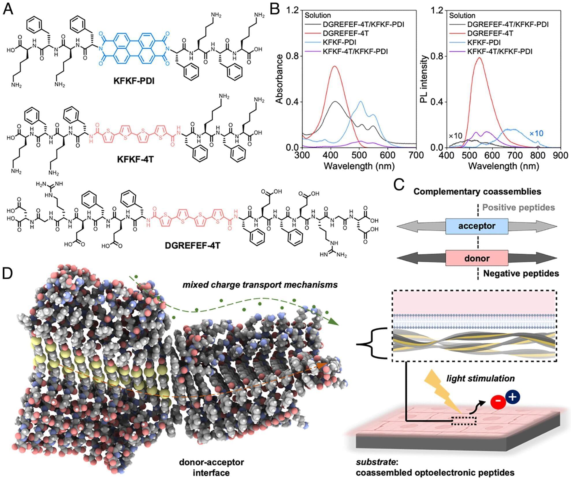
Toggle light / dark theme

In order for immune cells to do their job, they need to know against whom they should direct their attack. Research teams at the University of Würzburg have identified new details in this process.

As complicated as their name is, they are important for the human organism in the fight against pathogens and cancer: Vγ9Vδ2 T cells are part of the immune system and, as a subgroup of white blood cells, fight cells and cells infected with pathogens. They recognize their potential victims by their altered cell metabolism.

Research teams from the University of Würzburg and the University Hospital of Würzburg, together with groups in Hamburg, Freiburg, Great Britain and the U.S., have now gained new insights into how these cells manage to look inside the cell. Thomas Herrmann, Professor of Immunogenetics at the Institute of Virology and Immunobiology and his colleague Dr. Mohindar Karunakaran at Julius-Maximilians-Universität Würzburg (JMU), were responsible for the study published in the journal Nature Communications.

In this episode, Dr. David Sinclair and co-host Matthew LaPlante discuss why we age. In doing so, they discuss organisms that have extreme longevity, the genes that control aging (mTOR, AMPK, Sirtuins), the role of sirtuin proteins as epigenetic regulators of aging, the process of “ex-differentiation” in which cells begin to lose their identity, and how all of this makes up the “Information Theory of Aging”, and the difference between “biological age” and “chronological age” and how we can measure biological age through DNA methylation clocks. #Aging #DavidSinclair #Longevity

The advancement of higher cognitive abilities in humans is predominantly associated with the growth of the neocortex, a brain area key to conscious thinking, movement, and sensory perception. Researchers are increasingly realizing, however, that the “little brain” or cerebellum also expanded during evolution and probably contributes to the capacities unique to humans, explains Prof. Henrik Kaessmann from the Center for Molecular Biology of Heidelberg University.

His research team has – together with Prof. Dr Stefan Pfister from the Hopp Children’s Cancer Center Heidelberg – generated comprehensive genetic maps of the development of cells in the cerebella of humans, mice, and opossums. Comparisons of these data reveal both ancestral and species-specific cellular and molecular characteristics of cerebellum development spanning over 160 million years of mammalian evolution.

Genome and Structure:

HIV’s genome is a 9.7 kb linear positive-sense ssRNA.1 There is a m7G-cap (specifically the standard eukaryotic m7GpppG as added by the host’s enzymes) at the 5’ end of the genome and a poly-A tail at the 3’ end of the genome.2 The genome also has a 5’-LTR and 3’-LTR (long terminal repeats) that aid its integration into the host genome after reverse transcription, that facilitate HIV genetic regulation, and that play a variety of other important functional roles. In particular, it should be noted that the integrated 5’UTR contains the HIV promoter called U3.3,4

HIV’s genome translates three polyproteins (as well as several accessory proteins). The Gag polyprotein contains the HIV structural proteins. The Gag-Pol polyprotein contains (within its Pol component) the enzymes viral protease, reverse transcriptase, and integrase. The Gag-Pol polyprotein is produced via a −1 ribosomal frameshift at the end of Gag translation. Because of the lower efficiency of this frameshift, Gag-Pol is synthesized 20-fold less frequently than Gag.5 The frameshift’s mechanism depends upon a slippery heptanucleotide sequence UUUUUUA and a downstream RNA secondary structure called the frameshift stimulatory signal (FSS).6 This FSS controls the efficiency of the frameshift process.

An MIT study suggests 3D folding of the genome is key to cells’ ability to store and pass on “memories” of which genes they should express.


Every cell in the human body contains the same genetic instructions, encoded in its DNA. However, out of about 30,000 genes, each cell expresses only those genes that it needs to become a nerve cell, immune cell, or any of the other hundreds of cell types in the body.

Each cell’s fate is largely determined by chemical modifications to the proteins that decorate its DNA; these modification in turn control which genes get turned on or off. When cells copy their DNA to divide, however, they lose half of these modifications, leaving the question: How do cells maintain the memory of what kind of cell they are supposed to be?

A new MIT study proposes a theoretical model that helps explain how these memories are passed from generation to generation when cells divide. The research team suggests that within each cell’s nucleus, the 3D folding of its genome determines which parts of the genome will be marked by these chemical modifications. After a cell copies its DNA, the marks are partially lost, but the 3D folding allows the cell to easily restore the chemical marks needed to maintain its identity. And each time a cell divides, chemical marks allow a cell to restore its 3D folding of its genome. This way, by juggling the memory between 3D folding and the marks, the memory can be preserved over hundreds of cell divisions.

Researchers at the Hubrecht Institute have laid the foundation for the development of a gene therapy for the genetic heart disease arrhythmogenic cardiomyopathy (ACM). Their approach, based on replacement of the PKP2 gene, led to significant structural and functional improvements in laboratory models of the disease.

The study by the group of Eva van Rooij was published on 7 December 2023 in Nature Cardiovascular Research. Multiple will start in 2024 in the United States to explore the clinical potential of this approach in ACM patients with PKP2 mutations.

ACM is a genetic disease that affects 1 in 2,000 to 1 in 5,000 people worldwide. It is characterized by arrhythmias and can lead to sudden cardiac arrest. Current treatment of the disease usually consists of antiarrhythmic drugs and implantable cardioverter-defibrillators (ICDs), which are focused solely on treating the symptoms rather than targeting the root of the problem.

An international team led by researchers at the University of Toronto has uncovered over 100 genes that are common to primate brains but have undergone evolutionary divergence only in humans—and which could be a source of our unique cognitive ability.

The researchers, led by Associate Professor Jesse Gillis from the Donnelly Center for Cellular and Biomolecular Research and the department of physiology at U of T’s Temerty Faculty of Medicine, found the genes are expressed differently in the brains of humans compared to four of our relatives—chimpanzees, gorillas, macaques and marmosets.

The findings, published in Nature Ecology & Evolution, suggest that reduced , or tolerance to loss-of-function mutations, may have allowed the genes to take on higher-level cognitive capacity. The study is part of the Human Cell Atlas, a global initiative to map all to better understand health and disease.

On the internet, nothing is safe — not even your DNA, apparently.

That’s the dystopian lesson from the commercial genetic testing company 23andMe, which disclosed on Friday in a regulatory filing that hackers managed to access information on about 14,000 users or 0.1 percent of its customer user base.

But the problem goes beyond this relatively small number of people. Because the website allows users to share DNA information with other users in order to find relatives, the true number impacted is orders of magnitude larger — with about 6.9 million customers having their personal information compromised, according to TechCrunch. Big yikes on that figure, because it affects something like half of the 14 million users at 23andMe.