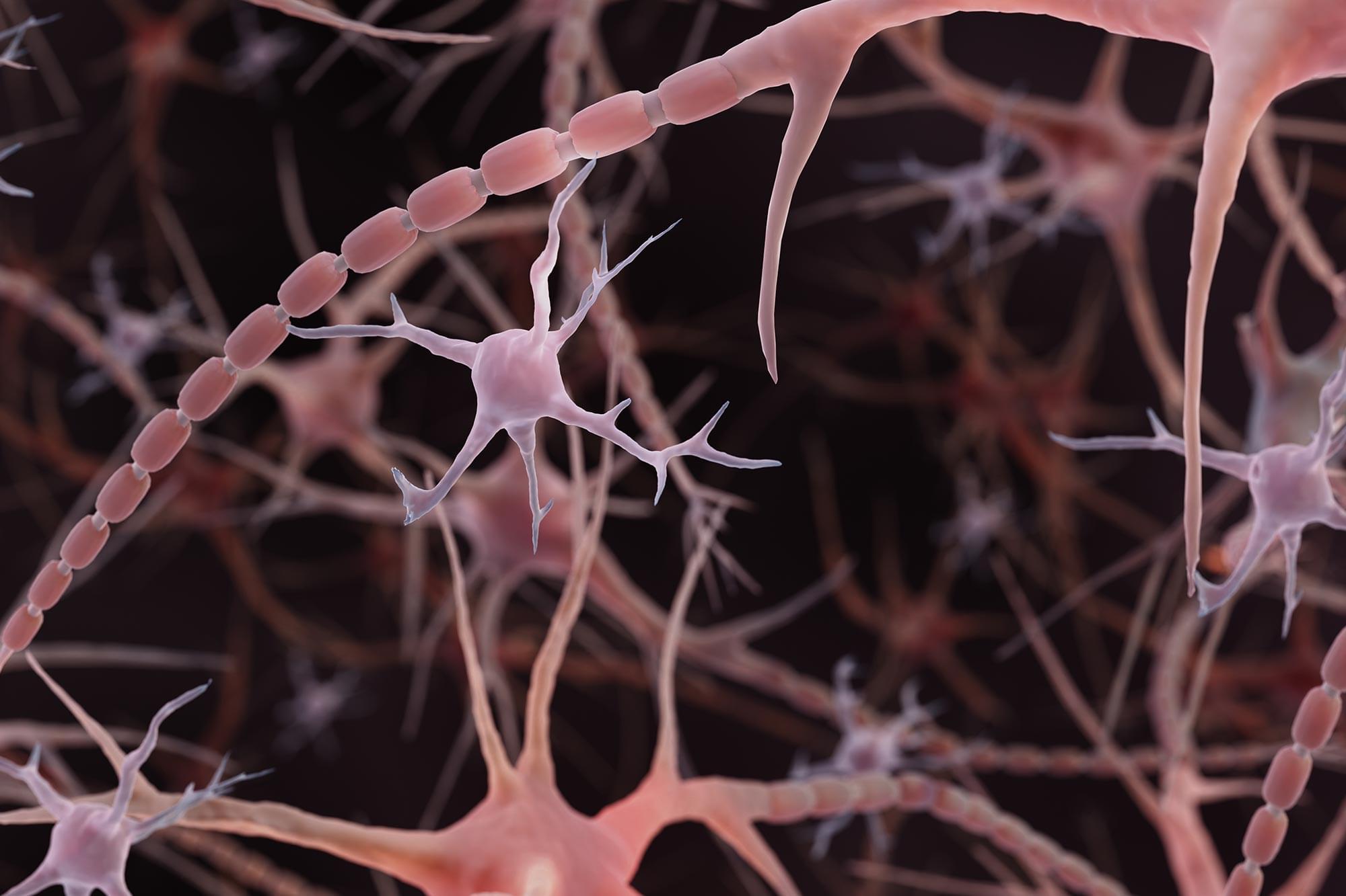
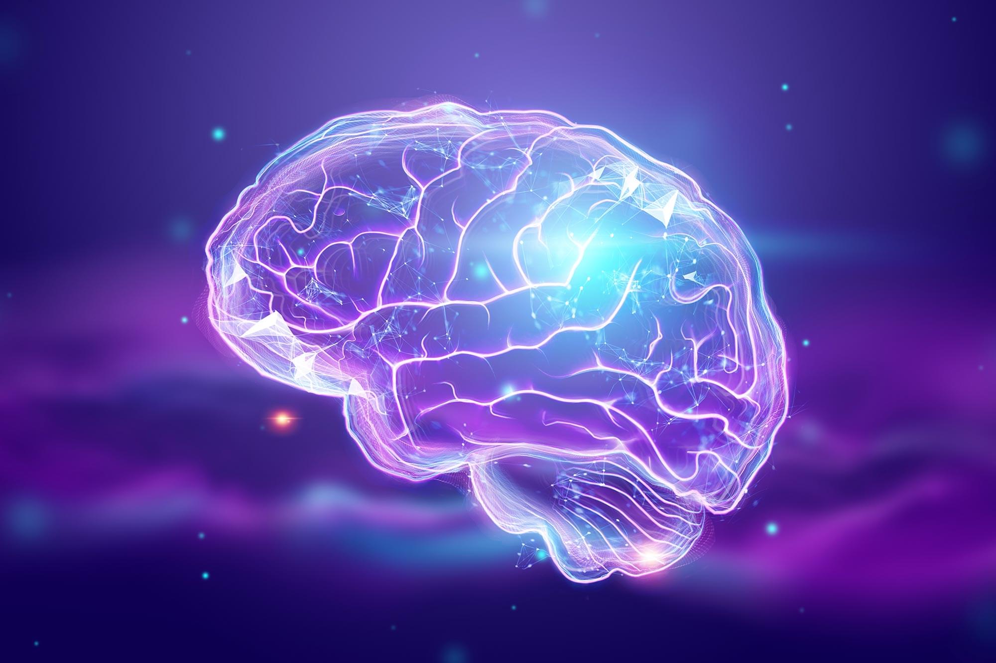
A newly identified brain receptor could supercharge immune cells to fight Alzheimer’s and protect memory.

Persistent genomic instability compromises cellular viability while also triggers non-cell-autonomous responses that drive dysfunction across tissues, contributing to aging. Recent evidence suggests that DNA damage activates secretory programs, including the release of inflammatory cytokines, damage-associated molecular patterns, and extracellular vesicles, that reshape immune homeostasis, stem cell function, and metabolic balance. Although these responses may initially support tissue integrity and organismal survival, their chronic activation has been associated with tissue degenerative changes and systemic decline. Here, we discuss how nuclear DNA damage responses trigger the activation of cytoplasmic sensing pathways, promote secretory phenotypes, and affect organismal physiology. Targeting DNA damage-driven mechanisms may help buffer harmful systemic responses while preserving regeneration and immune surveillance, offering new ways to delay aging-related decline.
© 2025 The Author(s). BioEssays published by Wiley‐VCH GmbH.
Research led by Rutgers suggests there could be significant new possibilities for treating neurodegenerative diseases and brain injuries.
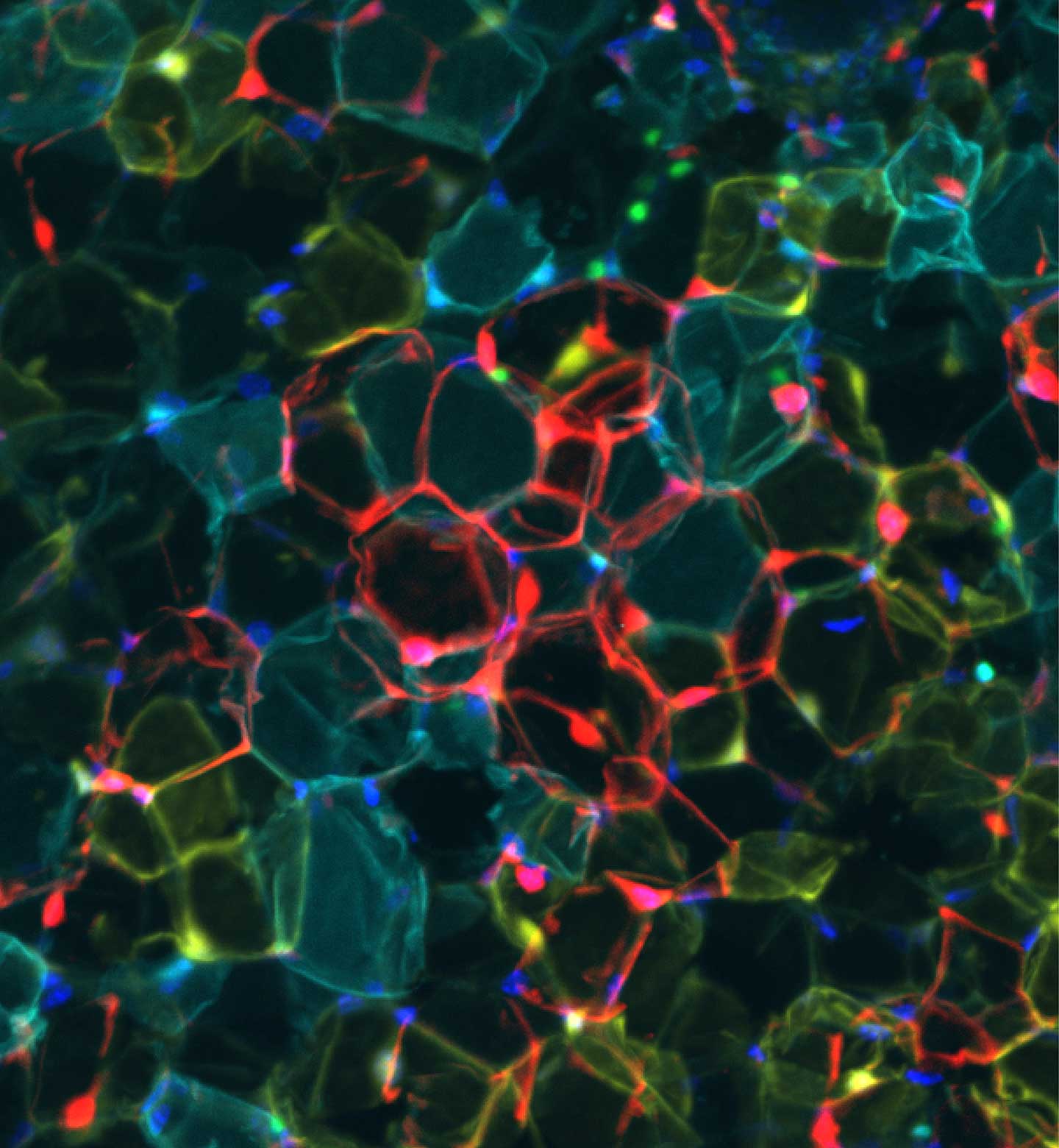
Researchers have uncovered how a specific protein supports the stability of connections between brain cells, which are essential for learning and memory.
According to the scientists, their findings, published in the journal Science Advances.
