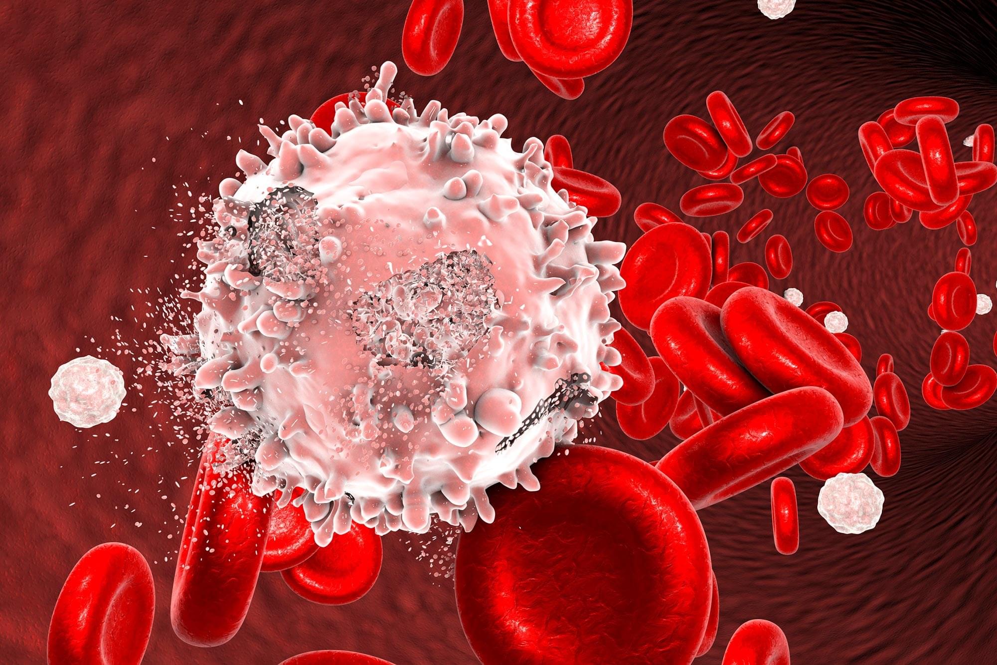
Marginal zone lymphoma treatment evolves with promising CAR T-cell therapies, addressing unmet needs in aggressive cases and improving patient outcomes.
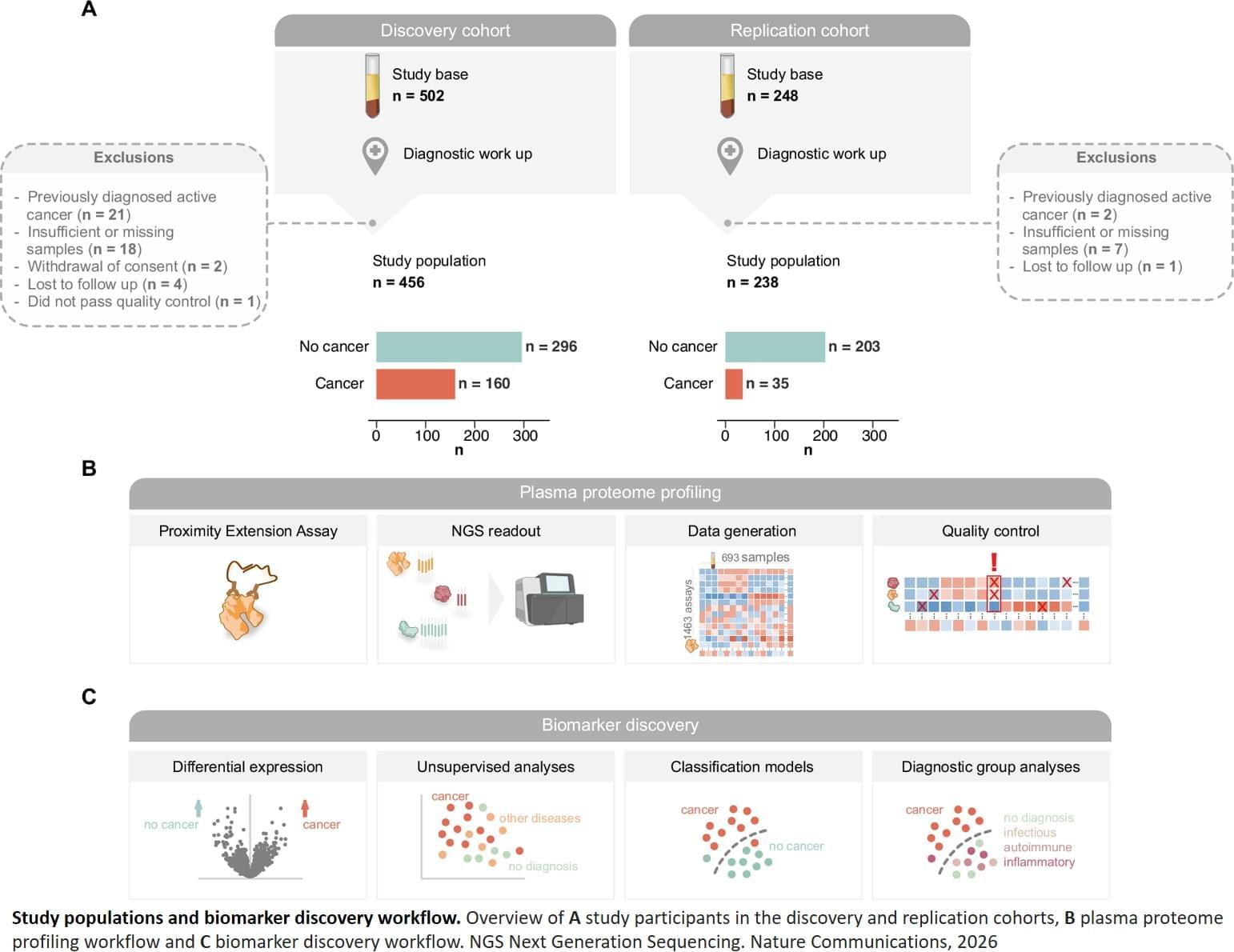
The researchers then developed a model that can distinguish patients with cancer from those with other conditions, such as inflammatory, autoimmune or infectious diseases, with high precision.
“A particular strength of the study is that the control group consisted largely of patients with other serious conditions that can cause symptoms similar to cancer,” says the principal investigator for the study. “This reflects the clinical reality, where patients with non-specific symptoms are often difficult to assess.”
The researchers emphasise that the method should not replace imaging diagnostics or biopsies, but rather serve as a support for prioritising which patients should be investigated further. ScienceMission sciencenewshighlights.
A simple blood test can help detect cancer in patients with non-specific symptoms such as fatigue, pain or weight loss. This is according to a study published in Nature Communications.
When patients seek care for non-specific symptoms such as fatigue, pain or weight loss, it is often difficult to determine whether the cause is cancer, another serious condition or something completely harmless. In a new study, researchers have investigated whether proteins in the blood can provide early clues.
The study analysed blood samples from nearly 700 patient and the samples were taken before the diagnostic investigation began. Using proteomics, a method for large-scale protein analysis, the levels of 1,463 different proteins in plasma were measured. The researchers identified a specific combination of proteins, known as a protein signature, that could be linked to a cancer diagnosis.
Schematic overview of the relationship between sinusitis, otitis media and/or mastoiditis, intracranial infection, and septic CVST. Management strategies include surgical washout of the infection, parenteral antibiotics, and hydration, although the role of anticoagulation remains controversial.
Septic cerebral venous sinus thrombosis (CVST) is a recognized complication of pediatric sinogenic and otogenic intracranial infections. The optimal treatment paradigm remains controversial. Proponents of anticoagulation highlight its role in preventing thrombus propagation and promoting recanalization, while others cite the risk of hemorrhagic complications, especially after a neurosurgical procedure for an epidural abscess or subdural empyema. Here, the authors investigated the diagnosis, management, and outcomes of pediatric patients with sinogenic or otogenic intracranial infections and a septic CVST.
All patients 21 years of age or younger, who presented with an intracranial infection in the setting of sinusitis or otitis media and who underwent neurosurgical treatment at Connecticut Children’s, Rady Children’s Hospital–San Diego, or Ann and Robert H. Lurie Children’s Hospital of Chicago from March 2015 to March 2023, were retrospectively reviewed. Demographic, clinical, and radiological data were systematically collated.
Ninety-six patients were treated for sinusitis-related and/or otitis media–related intracranial infections during the study period, 15 (15.6%) of whom were diagnosed with a CVST. Of the 60 patients who presented prior to the COVID-19 pandemic, 6 (10.0%) were diagnosed with a septic CVST, whereas of the 36 who presented during the COVID-19 pandemic, 9 (25.0%) had a septic CVST (p = 0.050). The superior sagittal sinus was involved in 12 (80.0%) patients and the transverse and/or sigmoid sinuses in 4 (26.7%). Only 1 (6.7%) patient had a fully occlusive thrombus. Of the 15 patients with a septic CVST, 11 (73.3%) were initiated on anticoagulation at a median interval of 4 (IQR 3–5) days from the most recent neurosurgical procedure. Five (45.5%) patients who underwent anticoagulation demonstrated complete recanalization on follow-up imaging, and 4 (36.4%) had partial recanalization. Three (75.0%) patients who did not undergo anticoagulation demonstrated complete recanalization, and 1 (25.
Finding evidence of is fraught, whether in a comatose patient, an animal, or a neural net.
A hidden immune cascade linking the gut and bone marrow may explain how IBD turns inflammation into colon cancer.
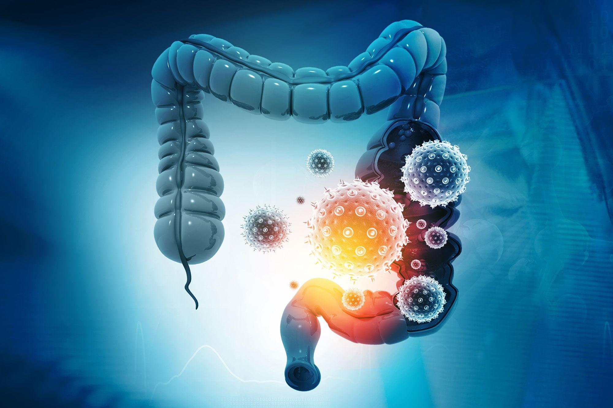
Scientists at Weill Cornell Medicine have identified a complex immune process in the gut that may help explain why people with inflammatory bowel disease (IBD) face a much higher risk of colorectal cancer. The preclinical study shows how a specific immune signal can trigger a wave of white blood cells from the bone marrow into the gut, creating conditions that support tumor growth. The findings also suggest new approaches for detecting disease activity, tracking risk, and developing future treatments.
The role of TL1A in gut inflammation.
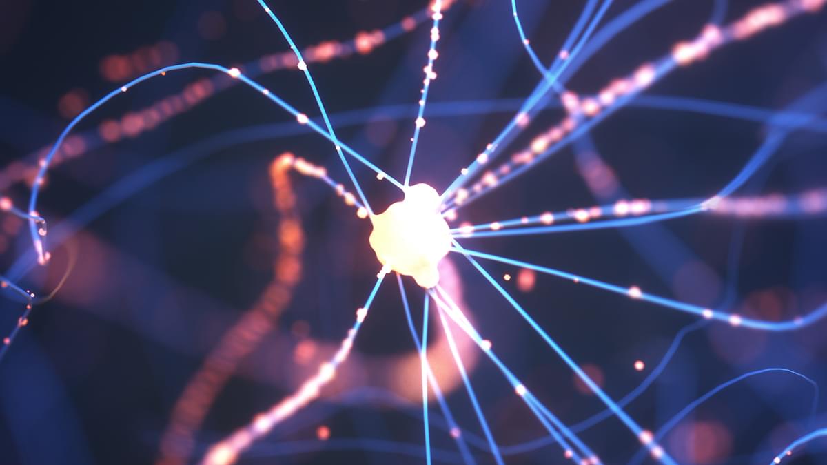
An investigation by researchers from Case Western Reserve University School of Medicine in the US has filled in a missing link between the toxic build-up of proteins in the neurodegenerative condition Parkinson’s disease and the death of critical brain cells.
The result of three years of research, the discovery connects alpha-synuclein proteins to a breakdown in mitochondrial function, both previously linked to Parkinson’s.
“We’ve uncovered a harmful interaction between proteins that damages the brain’s cellular powerhouses, called mitochondria,” says neuroscientist Xin Qi.
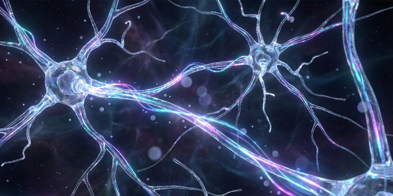
In the group treated with psilocybin, adapting neurons sat at a resting voltage that was closer to the threshold for firing. This state is known as depolarization. It means the cells are primed to activate more easily. The bursting neurons in psilocybin-treated rats also showed increased excitability. They required less input to trigger a signal and fired at faster rates than neurons in untreated rats.
The rats treated with 25CN-NBOH also exhibited functional changes, though the specific electrical alterations differed slightly from the psilocybin group. For instance, the bursting neurons in this group were not as easily triggered as those in the psilocybin group. However, the overall pattern confirmed that the drug had induced a lasting shift in neuronal function.
These electrophysiological findings provide a potential explanation for the behavioral results. While the physical branches of the neurons may have pruned back to normal levels, the cells “remembered” the treatment through altered electrical tuning. This functional shift allows the neural circuits to operate differently long after the drug has left the body.