Toggle light / dark theme

The ‘silent’ brain cells that shape our behaviour, memory and health

Researchers peered through microscopes, hooked up electrodes, and built entire careers around one cell type: neurons. These electrically active cells were clearly the brain’s protagonists, zipping signals through our heads at lightning speed to create thoughts, memories, and movements. Everything else—especially the star-shaped cells called astrocytes that outnumber neurons—was dismissed as mere scaffolding. Glial cells, they were called: “glue.”

Inbal Goshen, a memory researcher at Hebrew University of Jerusalem, remembers feeling like an outsider when she started investigating astrocytes in the early 2010s. “Oh, that’s the weird one who works on astrocytes,” she imagined colleagues whispering at conferences. The skepticism was palpable. Yet new molecular tools had finally given her a way to peek into these mysterious cells, and what she found was too intriguing to ignore.

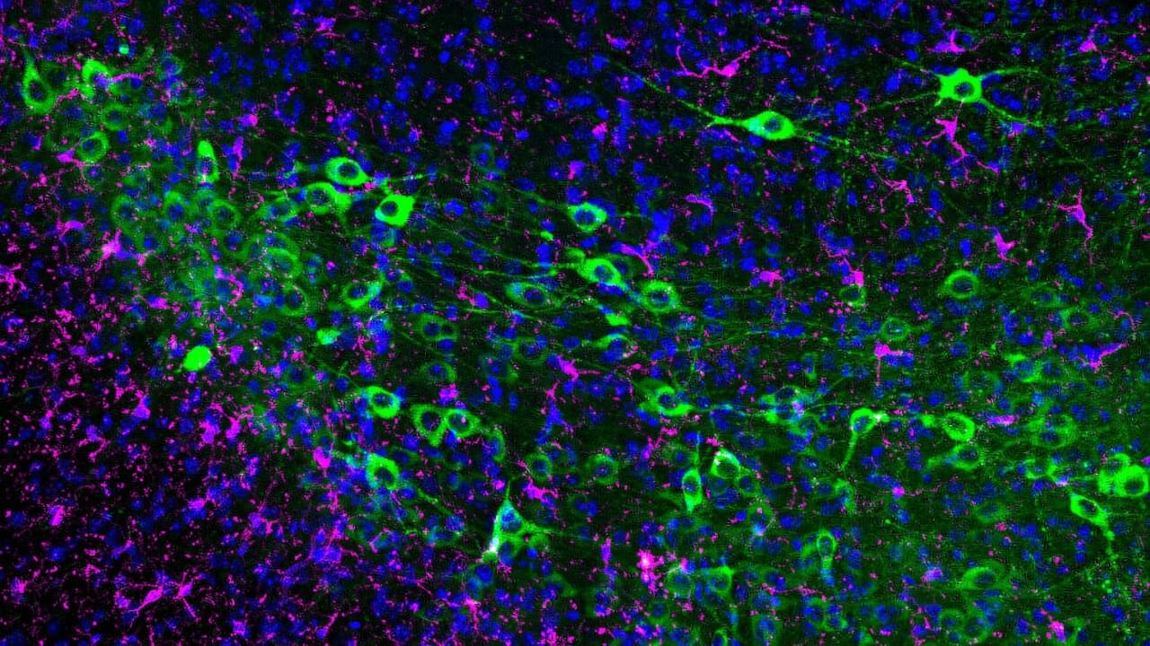
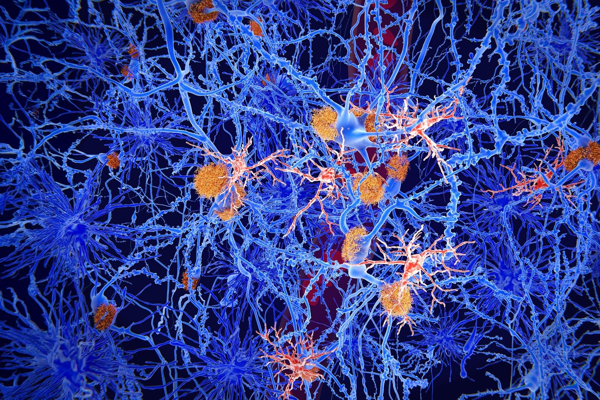
Unlike neurons, astrocytes don’t fire electrical signals. They were “electrically silent,” which is why they’d been ignored. But they were whispering in another language entirely: calcium. Using advanced imaging, researchers discovered that astrocytes communicate through slow, rhythmic waves of calcium signals—more like a gentle tide than neuron’s lightning strike. And their reach is astonishing: a single human astrocyte can touch up to two million synapses, the junctions where neurons meet. Their bushy tendrils fill every crevice of the brain, each cell nestling against neurons and blood vessels, creating an intimate, three-way relationship.

Memory research revealed another layer. Goshen’s team watched astrocytes in mice navigating toward water rewards. As the animals approached familiar prize locations, astrocyte activity slowly ramped up—but showed no response in new environments. The cells were encoding spatial memories, not just supporting them. Other labs found that astrocytes help stabilize and recall fear memories, their slow calcium signals perfectly suited to bridge the gap between learning something and remembering it days later. As neuroscientist Jun Nagai describes it, “Think of them as the brain’s long-exposure camera: they capture the trace of meaningful events that might otherwise fade too fast.”


Astrocytes make up one-quarter of the brain, but researchers are only now realizing their true value.

Pegcetacoplan—the ‘closest thing to a cure’ for rare, severe kidney disease

A rare and life-threatening kidney disease in children finally has an effective therapy, thanks in large part to pioneering research and clinical leadership from University of Iowa Health Care Stead Family Children’s Hospital.

The disease, known as C3 glomerulopathy (C3G), is an ultra-rare condition that primarily affects children and young adults. Only around 5,000 Americans have C3G, which causes progressive kidney damage, with more than half of patients reaching end-stage kidney failure within a decade of diagnosis.

Unlike previous treatments for C3G that aimed to alleviate the damaging inflammatory process of the disease, the new, first-of-its-kind drug directly targets the root cause of C3G dysfunction in the body’s complement system, a part of the immune response.

Abstract: 1 Institut de Neurociències, and

1 Institut de Neurociències, and.

2Department of Cell Biology, Physiology and Immunology, Universitat Autònoma de Barcelona, Bellaterra, Barcelona Spain.

3Institute of Neuroscience, CSIC-UMH, Alicante, Spain.

4Neurodegenerative Diseases Research Group, Vall d’Hebron Research Institute-Network Center for Biomedical Research in Neurodegenerative Diseases (CIBERNED), Barcelona, Spain.

Organisms are musicians not machines

The living cell, once thought to be a precise molecular factory, is turning out to be more like an improvising jazz ensemble. The old dogma—one gene, one protein, one function—has collapsed. Molecular biologist Ewa Grzybowska argues that recent discoveries show that proteins can switch folds, shift shapes, or even remain gloriously unstructured, improvising their roles as they go. Genes are not blueprints but texts, open to continuous interpretation by cells. Life, it turns out, is not built like a machine but is instead fluid, improvisational, and brimming with creative possibility.

1. The old paradigms in biology

When Watson and Crick decoded DNA in 1953 and the mechanism of protein-making was discovered, we obtained an extraordinary tool to explain the inner workings of life. The basic principle of making one protein from one DNA template (gene) with the assistance of one messenger RNA (mRNA) and several well-defined amino-acid-transporting RNAs (tRNAs) was so successful that it has been enshrined in millions of textbooks and not questioned for a long time.

How the brain protects itself from Alzheimer’s disease

High levels of calcium are toxic to cells and contribute to loss of neurons in Alzheimer’s disease. A new study published in JCI Insight identifies a mechanism through which the young brain protects itself against high calcium levels, and it could help scientists learn how to protect the brain from this devastating neurodegenerative condition.

Glyoxalase 1 (GLO1) is a protein that plays an essential role in getting rid of toxic byproducts in cells. In the study, Yale School of Medicine (YSM) researchers discovered elevated GLO1 levels in the brains of animals with excessive levels of cellular calcium, finding that the brain increased GLO1 expression as a protective mechanism to mitigate the effects of the calcium dysregulation.

However, with advancing age, GLO1 activity declined, the researchers found, which may make the brain less resilient to neurodegeneration. The study could inform the development of therapeutics that target GLO1 and prevent neurodegeneration.

Cracking the code of Parkinson’s: How supercomputers are pointing to new treatments

More than 1 million Americans live with tremors, slowed movement and speech changes caused by Parkinson’s disease—a degenerative and currently incurable condition, according to the Parkinson’s Foundation and the Mayo Clinic. Beyond the emotional toll on patients and families, the disease also exerts a heavy financial burden. In California alone, researchers estimate that Parkinson’s costs the state more than 6 billion dollars in health care expenses and lost productivity.

Scientists have long sought to understand the deeper brain mechanisms driving Parkinson’s symptoms. One long-standing puzzle involved an unusual surge of brain activity known as beta waves—electrical oscillations around 15 Hertz observed in patients’ motor control centers. Now, thanks to supercomputing resources provided by the U.S. National Science Foundation’s ACCESS program, researchers may have finally discovered what causes these waves to spike.

Using ACCESS allocations on the Expanse system at the San Diego Supercomputer Center—part of UC San Diego’s new School of Computing, Information, and Data Sciences—researchers with the Aligning Science Across Parkinson’s (ASAP) Collaborative Research Network modeled how specific brain cells malfunction in Parkinson’s disease. Their findings could pave the way for more targeted treatments.

ALS treatment using RNA-based gene therapy

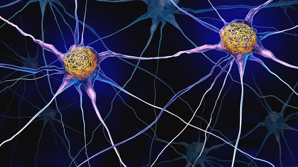
The current study was based on a feature of ALS discovered previously in the lab: toxic clusters (aggregates) of a protein called TDP-43 (usually responsible for regulating protein production at the site) form at the tip of the nerve, where it meets the muscle. To discover how these TDP-43 aggregates are formed, the researchers used a mouse model for ALS, tissues from ALS patients, and cultures of human stem cells.

The study found that muscle cells produce small RNA molecules called microRNA-126 and send them in vesicles, through the synapsis, to the tip of the nerve cell. The role of these molecules is to prevent the expression of the TDP-43 protein at the neuromuscular junction when it is not needed.

The author explains: “We discovered that in ALS, the muscle produces a smaller amount of microRNA-126, which leads to an excess of TDP-43. The excess protein forms toxic aggregates that attack molecules essential for functioning of the mitochondria — the nerve cell’s powerhouse. Damage to the mitochondria causes an energy deficit, gradually destroying motor neurons and leaving patients’ muscles paralyzed.”

The study further showed that when the amount of microRNA-126 is reduced, a process similar to ALS occurs, and the neurons are destroyed. Conversely, increasing the level of microRNA-126 in tissues taken from ALS patients and in ALS model mice led to a decrease in the levels of TDP-43, and the neurons stopped degenerating and even regenerated.

The researchers concluded that adding microRNA-126 rescues neurons damaged by ALS, prevents degeneration of the neuromuscular junction, and could serve as a basis for developing effective drugs for this currently incurable disease.


Researchers in this study opened a new avenue for treating the fatal degenerative disease ALS, considered incurable until now. The researchers identified a new molecular mechanism that plays a key role in the disease and were able to neutralize it through gene therapy.

/* */