A major UK study reveals four childhood factors that can accelerate organ aging decades later, and why education may protect longevity.

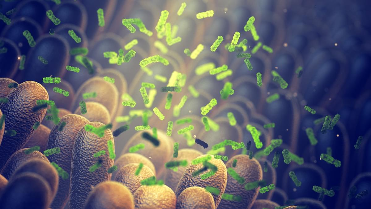
A compound produced by gut bacteria could play a vital role in managing and preventing type 2 diabetes, according to a study led by researchers from Imperial College London (ICL).
The small molecule, called trimethylamine (TMA), is a major type of bacterial metabolite – a class of chemicals produced naturally through processes of transforming nutrients into energy and building blocks.
Scientists have now found evidence in human cell models and lab mice that TMA could protect the body from some of the damage triggered by a high-fat diet. Specifically, it has the effect of dampening down inflammation and improving insulin response, both of which reduce the risk of type 2 diabetes.
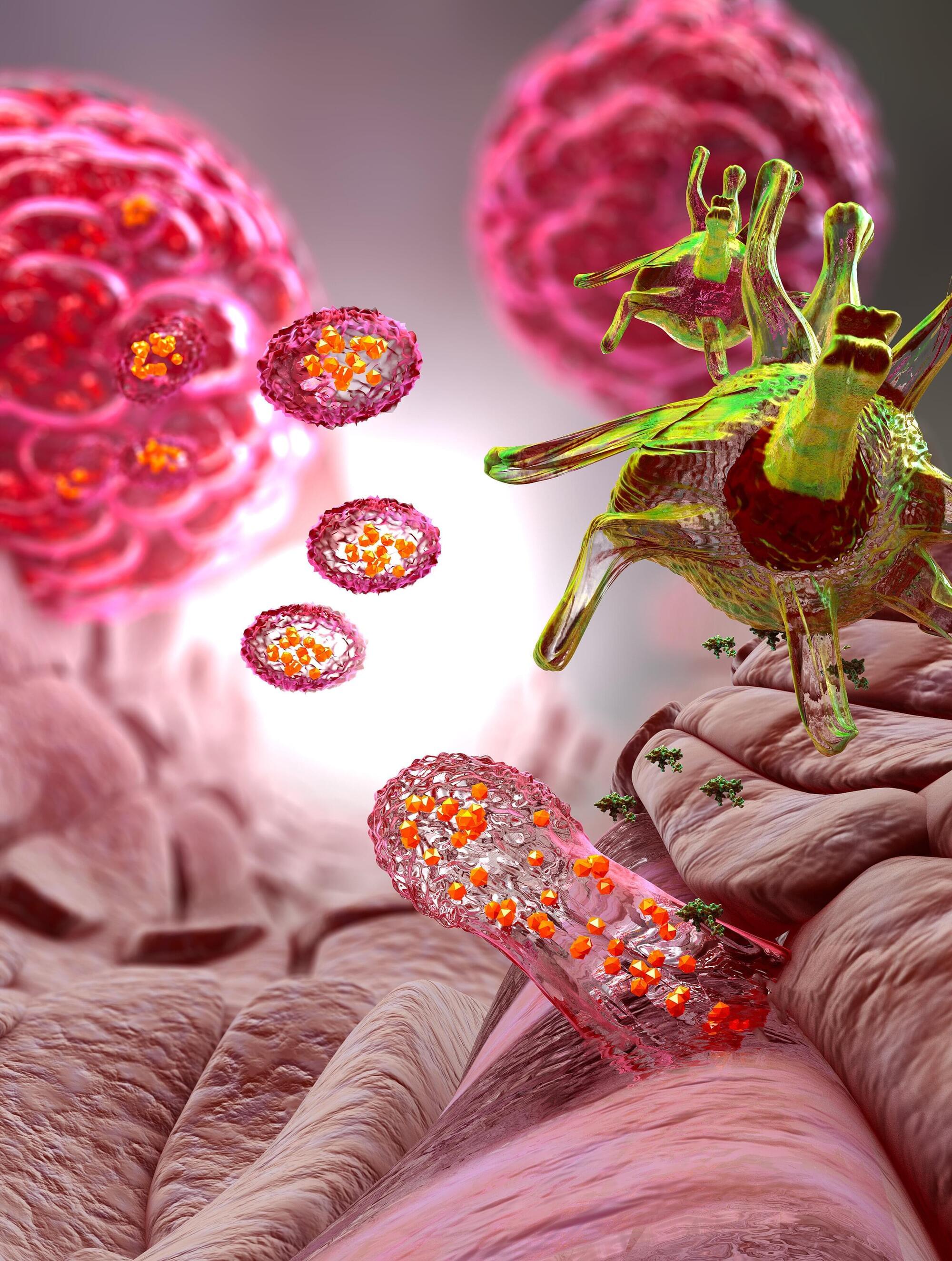
EPFL researchers have successfully engineered cells of the immune system to more effectively recognize cancer cells. The work, covered in two papers, turns the previously lab-based method into a full-blown immunotherapy strategy.
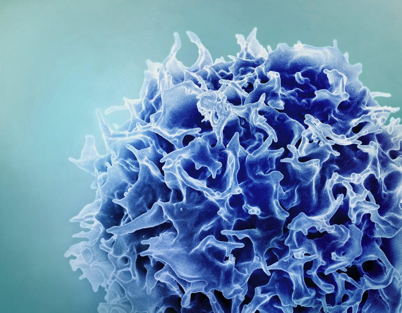
Cancer immunotherapy is a strategy that turns the patient’s own immune cells into a “search-and-destroy” force that attacks the tumor’s cells. The “search” immune cells are the dendritic cells, which collect and present identifying parts of the cancer cells (antigens) to the “destroy” part (T cells), the immune system’s killer cells.
The problem is that many tumors “learn” how to evade detection by the patient’s dendritic cells. Clinicians address this by collecting dendritic cells from the patient’s blood, loading them in the laboratory with tumor material—antigens that train dendritic cells to better identify the tumor—and then injecting them back into the patient.
Despite risks, results from both trials highlight the promise of one-and-done CAR T therapy for deadly blood cancers. But it’s still early days. Scientists need to carefully follow patients over years to understand how long upgraded T cells remain in the body and their effect on cancers.
And not all viral carriers are made the same. Lentiviruses, used in both studies, can tunnel into the human genome, causing DNA typos that potentially trigger secondary cancers. The durability of the therapy, its longevity, and immune side effects also need to be studied.
Kelonia is adding more patients to their trial, amid an increasingly competitive landscape. AstraZeneca has acquired EsoBiotec to bring its technology to market. AbbVie, a drug company in Illinois, is testing the delivery of gene-editing tools to T cells via fatty nanoparticles in clinical trials. And Kelonia is planning a second clinical trial with an initial 20 patients and 20 more in an expansion phase, none of whom responded to at least three previous treatments.
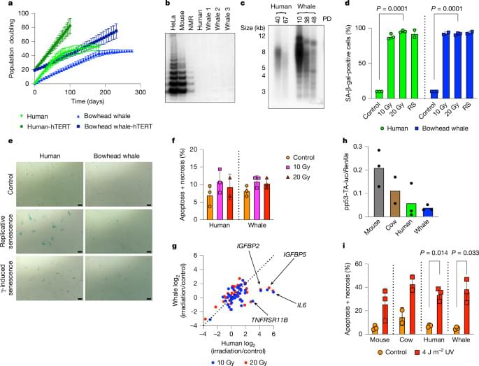
At more than 200 years, the maximum lifespan of the bowhead whale exceeds that of all other mammals. The bowhead is also the second-largest animal on Earth1, reaching over 80,000 kg. Despite its very large number of cells and long lifespan, the bowhead is not highly cancer-prone, an incongruity termed Peto’s paradox2.
Here, to understand the mechanisms that underlie the cancer resistance of the bowhead whale, we examined the number of oncogenic hits required for malignant transformation of whale primary fibroblasts. Unexpectedly, bowhead whale fibroblasts required fewer oncogenic hits to undergo malignant transformation than human fibroblasts. However, bowhead whale cells exhibited enhanced DNA double-strand break repair capacity and fidelity, and lower mutation rates than cells of other mammals. We found the cold-inducible RNA-binding protein CIRBP to be highly expressed in bowhead fibroblasts and tissues.
Bowhead whale CIRBP enhanced both non-homologous end joining and homologous recombination repair in human cells, reduced micronuclei formation, promoted DNA end protection, and stimulated end joining in vitro. CIRBP overexpression in Drosophila extended lifespan and improved resistance to irradiation. These findings provide evidence supporting the hypothesis that, rather than relying on additional tumour suppressor genes to prevent oncogenesis3,4,5, the bowhead whale maintains genome integrity through enhanced DNA repair. This strategy, which does not eliminate damaged cells but faithfully repairs them, may be contributing to the exceptional longevity and low cancer incidence in the bowhead whale.
Analysis of the longest-lived mammal, the bowhead whale, reveals an improved ability to repair DNA breaks, mediated by high levels of cold-inducible RNA-binding protein.
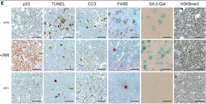
(Cell Reports 3, 1512–1525; May 30, 2013)
In the originally published paper, the CC3 panel for the +/+ sample in Figure 6E was inadvertently duplicated from the TUNEL panel of the +/RR sample during figure assembly. The authors have retrieved the correct CC3 image for the +/+ sample and assembled a corrected version of Figure 6E, which is provided below. The authors apologize for the error.

Reducing calorie intake helps cancer-fighting immune cells do their jobs more effectively, reports a study by Van Andel Institute scientists and collaborators. The findings lay the groundwork for developing dietary strategies to boost the effects of a powerful class of cancer immunotherapies.
“Growing evidence suggests dietary restriction has anti-cancer effects but the ‘why and how’ are not well understood. Our new study reveals one way this relationship may work: by providing T cells, the soldiers of the immune system, with the right mix of nutrients to more effectively fight cancer,” said Russell Jones, Ph.D., chair of VAI’s Department of Metabolism and Nutritional Programming and corresponding author of the study.
“Additional research is needed but we are hopeful these insights can inform evidence-based dietary guidelines to improve the effectiveness of immune-based cancer treatments.”
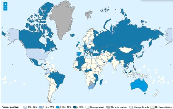
Seasonal influenza activity has increased globally in recent months, and influenza A(H3N2) viruses are predominant. This rise coincides with the onset of winter in the northern hemisphere. Epidemics and outbreaks of seasonal influenza and other circulating respiratory viruses can place significant pressure on healthcare systems. Although global activity remains within expected seasonal ranges, early increases and higher activity than typical at this time of year have been observed in some regions. Seasonal influenza could place significant pressure on healthcare systems even in non-temperate countries. Genetically drifted influenza A(H3N2) viruses, known as subclade K viruses, have been detected in many countries. While data on how well the vaccine works against clinical disease this season are still limited, vaccination is still expected to protect against severe illness and remains one of the most effective public health measures.
Surveillance
Due to the constantly evolving nature of influenza viruses, WHO continues to stress the importance of year-round global surveillance to detect and monitor virological, epidemiological and clinical changes associated with emerging or circulating influenza viruses that may affect human health and timely virus sharing for risk assessment. Countries are encouraged to remain vigilant to the threat of influenza viruses and review any unusual epidemiological patterns.
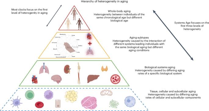
We developed a single blood-based methylation test that estimates biological aging across 11 physiological systems. This multisystem measure predicts mortality and health outcomes more precisely than existing epigenetic clocks, and reveals distinct aging patterns that could guide personalized gerotherapeutic and geroprotective interventions.