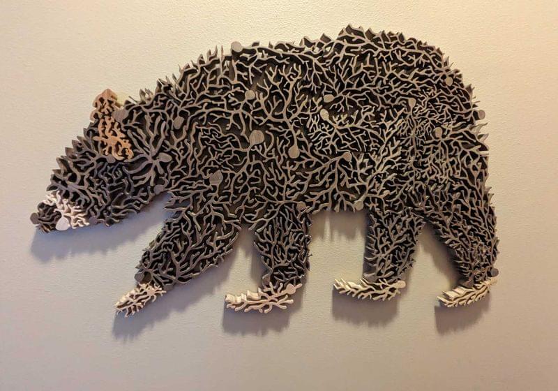
Researchers have created functional brain-like tissue without relying on any animal-derived materials, marking a major step toward more ethical and reproducible neurological research.
Researchers at the University of Eastern Finland and their international collaborators have identified key developmental and molecular differences between the two main subtypes of chronic lymphocytic leukemia, CLL. The findings, published in PLOS ONE, show that mutated and unmutated forms of CLL may originate from distinct stages of B cell development, offering new insight into disease mechanisms and biomarker discovery.
CLL, the most common leukemia in adults, is characterized by disruption of the peripheral immune system through the accumulation of abnormal B-lymphocytes. CLL is divided into mutated (M-CLL) and unmutated (UM-CLL) subtypes based on the mutation frequency of the immunoglobulin heavy chain variable region in B cells. UM-CLL is more aggressive and tends to have a worse prognosis than M-CLL. The research team performed a meta-analysis of transcriptomic data from 116 patients and healthy donor B cells to explore the origins of these subtypes.
B cells go through different developmental stages in bone marrow and in lymphatic tissue germinal centers. They are classified into different subtypes depending on their maturation and function, such as memory or plasma cells. The results revealed that M-CLL resembles germinal center–dependent memory B cell subtype, CD27bright memory B cells, while UM-CLL reflects an earlier intermediary germinal center stage, possibly explaining their differences in mutation levels and clinical behavior.
Pre-order LymeChart: https://LymeChart.ai/?utm_term=ryan5
(LymeChart is a product of linkaChart Medical)
Neura Pod is a series covering topics related to Neuralink, Inc. Topics such as brain-machine interfaces, brain injuries, and artificial intelligence will be explored. Host Ryan Tanaka synthesizes information, shares the latest updates, and conducts interviews to easily learn about Neuralink and its future.
Sign up for Neuralink’s Patient Registry: https://neuralink.com/trials/
Join the Neuralink team: https://neuralink.com/careers/
X: https://www.x.com/ryantanaka3/
Support: https://www.patreon.com/neurapod/
Join our LONGEVITY and ANTI-AGING Skool Community: https://www.skool.com/youthspan-society-9710/
Timestamps:
00:00 Intro.
00:41 DNA damage and aging.
01:37 Why bowhead whales live so long.
03:13 Cold shock proteins and lifespan.
04:43 Body temperature and longevity.
06:50 Acute cold exposure benefits.
08:10 Takeaway.
100 Health Biomarkers Ranked: https://youtu.be/SgKp5mm0ALI?si=M7YkYo6Lelci7kOQ
Start rewinding your biological clock: https://www.siimland.co/course.
P.S. This is not professional medical advice and should not be taken as such. The creator of this video is not held accountable for your health. Consult your doctor first.
New research finds that dying cells leave a “footprint of death” that guides immune responses — but viruses like influenza can exploit this signaling. The discovery, published in Nature Communications, offers new insight into cell death, viral transmission, and potential drug targets.
New insights into the aftermath of cell death might ultimately inform drug development.
Multicellular organisms (animals, plants, humans) all have the ability to methylate the cytosine base in their DNA. This process, a type of epigenetic modification, plays an important role in conditions such as cancer and processes such as aging.
In a paper appearing in Nature Genetics, researchers discover that in more “primitive” unicellular organisms, both the adenine and the cytosine bases are methylated. This would suggest that in some ways, these unicellular organisms are more complex than their multicellular peers.
The team also found that methylation of the adenine base was, in the case of many of these unicellular organisms, vital for controlling which genes are switched on, which is important for their viability.
Researchers at the University of Tokyo have built a microscope that can detect a signal over an intensity range 14 times wider than conventional microscopes. Moreover, the observations are made label-free, that is, without the use of additional dyes.
This means the method is gentle on cells and adequate for long-term observations, holding potential for testing and quality control applications in the pharmaceutical and biotechnology industries. The findings are published in the journal Nature Communications.
Microscopes have played a pivotal role in the development of science since the 16th century. However, progress has required not only more sensitive and accurate equipment and analysis, but also more specialized ones. Therefore, modern, cutting-edge techniques have had to straddle trade-offs.
“Our oral health is connected to our general health,” said Dr Sadia Niazi, a senior clinical lecturer in endodontology at King’s College London. “We should never look at our teeth or dental disease as a separate entity.”
Root canal treatment is one of the most common – and perhaps most feared – dental procedures, though much of the anxiety derives from myths and misconceptions that hark back to the days of poor anaesthetics. The treatment is performed to treat infection or damage to the tooth’s pulp, the soft inner tissue of a tooth that contains nerves, blood vessels and connective tissue.
Read More
If a looming root canal treatment is putting a dampener on the week, take heart: having the procedure can drive health benefits that are felt throughout the body, according to research.
Patients who were successfully treated for root canal infections saw their blood sugar levels fall significantly over two years, suggesting that ridding the body of the problematic bacteria could help protect against type 2 diabetes.
Dentists also saw improvements in patients’ blood cholesterol and fatty acid levels, both of which are associated with heart health. Yet more benefits were seen around inflammation, a driver for cardiovascular disease and other chronic conditions.