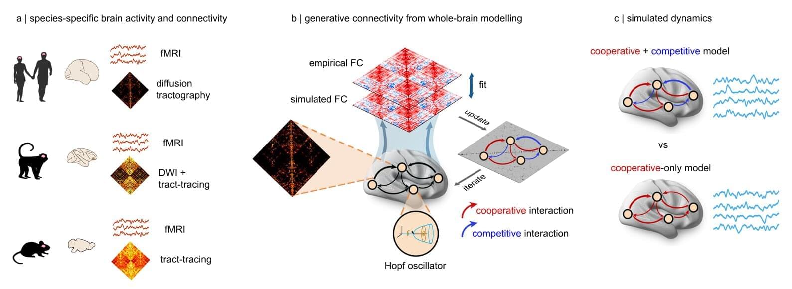
Toggle light / dark theme

A recent study investigates the relationship between exercise and the expression of MYC in skeletal muscles over time, revealing that even minimal doses can promote muscle growth without physical activity.

Researchers have long known that there is a relationship between the cancer-associated gene MYC (pronounced “Mick”) and exercise adaptation. When human muscles are exercised, MYC is found to increase transiently in abundance over 24 hours. But as we age, the MYC response to exercise is blunted, perhaps explaining a reduced ability to recover from exercise and maintain or gain muscle.

Knowing the precise mechanisms by which MYC drives muscle growth could prove instrumental in creating therapies that reduce muscle loss from aging, potentially improving independence, mobility, and health.

A new study finds tweaking part of the H5N1 virus infecting dairy cows in a single spot could allow it to better attach to human cell receptors, raising concerns it could transmit more easily between people.

By Lauren J. Young

Scientists have discovered that H5N1, the strain of highly pathogenic avian influenza virus currently spreading in U.S. dairy cows, only needs a single mutation to readily latch on to human cells found in the upper airway. The findings, published today in Science, illustrate a potential one-step path for the virus to become more effective at human transmission—and could have major implications for a new pandemic if such a mutation were to become widespread in nature.

Guest Editors: Prof. Robert Mannel, MD, University of Oklahoma HSC Prof. Judith Campisi, PhD, Buck Institute for Research on Aging Prof. Balazs Gyorffy, MD, PhD, Semmelweis University Prof. Anna Csiszar, MD, PhD, University of Oklahoma HSC Prof. Peter Bai, PhD, University of Debrecen.

The field of geroscience, focusing on the biology of aging, has revealed fascinating insights into the intricate relationship between aging and cancer. As the incidence of numerous cancer types exponentially increases with advancing age, understanding the cellular and molecular mechanisms underlying aging becomes crucial in deciphering the genesis and progression of cancer. We invite researchers to submit papers that shed light on specific mechanisms of aging that play pivotal roles in the development and progression of cancer, serve as targets for cancer treatments and contribute to the side effects of cancer therapies. Additionally, we are also interested in exploring the potential of aging-related biomarkers, including gene expression profiles associated with aging processes, as predictors of cancer survival.

📢New in GeroScience by Springer Nature.

Senescence cell signature associated with poor prognosis, epithelial–mesenchymalion, solid histology, and spread through air spaces in lung adenocarcinoma by Young Wha Koh, Jae-Ho Han, Seokjin Haam & Hyun Woo Lee.


Cellular senescence is involved in critical processes in tumor progression. Despite this potential relationship, the relationship between tumor cell senescence, prognostic significance, spread through air spaces (STAS), and tumor histology has not been investigated in lung adenocarcinoma (LUAD). We used the LUAD PanCancer Atlas dataset to assess senescence cell signature (SCS) based on the SenMayo gene set. We examined the relationship between SCS, prognostic significance, STAS, and tumor histology. This relationship was confirmed in independent LUAD datasets by validation using immunohistochemical senescence markers. In the LUAD PanCancer Atlas dataset, patients with high SCS expression had a higher prevalence of solid histology and STAS patterns than those with low SCS expression.

Researchers at the University of Adelaide, as part of an international team, have developed an approach that makes advanced microscopy possible through an optical fiber thinner than a human hair.

“Recent advances in optics have made it possible to controllably deliver light through extremely thin optical fibers, but delivering more complicated light patterns that are needed to perform advanced microscopy has eluded researchers until now,” said Dr. Ralf Mouthaan from the University of Adelaide’s Center of Light for Life, who undertook the project.

With a footprint far smaller than any other fiber imaging device, this will enable microscope images to be collected from previously inaccessible parts of the human body, while minimizing associated tissue damage.

In a recent study published in Nature Communications, a team of researchers at the Carl R. Woese Institute for Genomic Biology reports a new, robust computational toolset to extract biological relationships from large transcriptomics datasets. These efforts will help scientists better investigate cellular processes.

Living organisms are governed by their genome—an instruction manual written in the language of DNA that dictates how an organism grows, survives, and reproduces. By regulating the abundance of different RNA transcripts, cells control their protein expression level, thereby shaping their functions and responses to the environment.

Transcriptomics is the study of gene expression through cataloging the presence and abundance levels of active RNA transcripts generated from the genome under different conditions. Through the lens of RNA, transcriptomics technologies allow scientists to study the that enable life and cause disease, as well as assess the biological effects of therapeutics.

Unlocking Real-Time Inflammation Monitoring with Active-Reset Protein Sensors.

Imagine a tiny device that could continuously monitor your body’s inflammation levels, offering insights in real time to help manage diseases. While sensors for small molecules like glucose have existed for years, tracking proteins—a critical component in understanding inflammation—has been a major challenge. Proteins are present at much lower levels in the body, and traditional sensors struggle with slow response times due to their high-affinity binding to these molecules.

A team led by Zargartalebi has now overcome this barrier by introducing active-reset protein sensors. These sensors employ high-frequency oscillations of positive voltage to rapidly release bound protein molecules from their sensing electrodes. This breakthrough allows the sensors to reset in under a minute, enabling continuous tracking of protein levels.

The researchers incorporated these sensors into a microneedle device, which was successfully tested in mice to monitor cytokines—key markers of inflammation associated with diabetes. Unlike previous approaches, this method is not only simple to implement but can also be adapted to various sensor designs, potentially revolutionizing how we monitor and respond to conditions like chronic inflammation.

By bringing real-time protein monitoring closer to reality, active-reset sensors could pave the way for more dynamic and responsive healthcare, ensuring better disease management and prevention.

[#biosensors](https://www.facebook.com/hashtag/biosensors?__eep__=6&__cft__[0]=AZXpWTjqoXcElrcVMft-YBHF33SrUIfWG14SxebDv6jc7ayB9obklbDJ5BXDEcXMFhRK4gARwyDe24ko0Pjq0q5uSGpyaiia1YlxceB88HQ4AkLMRRKBaP_TJR0TQWWUQlYvZFrxBhxODGJhyWYo_RjJbdkcNE9A_kXuT2XJxXfpKD1LGhewSjGBaGndlInALDM&__tn__=*NK-R) [#invivo](https://www.facebook.com/hashtag/invivo?__eep__=6&__cft__[0]=AZXpWTjqoXcElrcVMft-YBHF33SrUIfWG14SxebDv6jc7ayB9obklbDJ5BXDEcXMFhRK4gARwyDe24ko0Pjq0q5uSGpyaiia1YlxceB88HQ4AkLMRRKBaP_TJR0TQWWUQlYvZFrxBhxODGJhyWYo_RjJbdkcNE9A_kXuT2XJxXfpKD1LGhewSjGBaGndlInALDM&__tn__=*NK-R) [#sensing](https://www.facebook.com/hashtag/sensing?__eep__=6&__cft__[0]=AZXpWTjqoXcElrcVMft-YBHF33SrUIfWG14SxebDv6jc7ayB9obklbDJ5BXDEcXMFhRK4gARwyDe24ko0Pjq0q5uSGpyaiia1YlxceB88HQ4AkLMRRKBaP_TJR0TQWWUQlYvZFrxBhxODGJhyWYo_RjJbdkcNE9A_kXuT2XJxXfpKD1LGhewSjGBaGndlInALDM&__tn__=*NK-R) [#inflammation](https://www.facebook.com/hashtag/inflammation?__eep__=6&__cft__[0]=AZXpWTjqoXcElrcVMft-YBHF33SrUIfWG14SxebDv6jc7ayB9obklbDJ5BXDEcXMFhRK4gARwyDe24ko0Pjq0q5uSGpyaiia1YlxceB88HQ4AkLMRRKBaP_TJR0TQWWUQlYvZFrxBhxODGJhyWYo_RjJbdkcNE9A_kXuT2XJxXfpKD1LGhewSjGBaGndlInALDM&__tn__=*NK-R) [#Microneedle](https://www.facebook.com/hashtag/microneedle?__eep__=6&__cft__[0]=AZXpWTjqoXcElrcVMft-YBHF33SrUIfWG14SxebDv6jc7ayB9obklbDJ5BXDEcXMFhRK4gARwyDe24ko0Pjq0q5uSGpyaiia1YlxceB88HQ4AkLMRRKBaP_TJR0TQWWUQlYvZFrxBhxODGJhyWYo_RjJbdkcNE9A_kXuT2XJxXfpKD1LGhewSjGBaGndlInALDM&__tn__=*NK-R)

[#regenerativesensors](https://www.facebook.com/hashtag/regenerativesensors?__eep__=6&__cft__[0]=AZXpWTjqoXcElrcVMft-YBHF33SrUIfWG14SxebDv6jc7ayB9obklbDJ5BXDEcXMFhRK4gARwyDe24ko0Pjq0q5uSGpyaiia1YlxceB88HQ4AkLMRRKBaP_TJR0TQWWUQlYvZFrxBhxODGJhyWYo_RjJbdkcNE9A_kXuT2XJxXfpKD1LGhewSjGBaGndlInALDM&__tn__=*NK-R) [#pointofcare](https://www.facebook.com/hashtag/pointofcare?__eep__=6&__cft__[0]=AZXpWTjqoXcElrcVMft-YBHF33SrUIfWG14SxebDv6jc7ayB9obklbDJ5BXDEcXMFhRK4gARwyDe24ko0Pjq0q5uSGpyaiia1YlxceB88HQ4AkLMRRKBaP_TJR0TQWWUQlYvZFrxBhxODGJhyWYo_RjJbdkcNE9A_kXuT2XJxXfpKD1LGhewSjGBaGndlInALDM&__tn__=*NK-R) [#diagnostics](https://www.facebook.com/hashtag/diagnostics?__eep__=6&__cft__[0]=AZXpWTjqoXcElrcVMft-YBHF33SrUIfWG14SxebDv6jc7ayB9obklbDJ5BXDEcXMFhRK4gARwyDe24ko0Pjq0q5uSGpyaiia1YlxceB88HQ4AkLMRRKBaP_TJR0TQWWUQlYvZFrxBhxODGJhyWYo_RjJbdkcNE9A_kXuT2XJxXfpKD1LGhewSjGBaGndlInALDM&__tn__=*NK-R) [#cytokines](https://www.facebook.com/hashtag/cytokines?__eep__=6&__cft__[0]=AZXpWTjqoXcElrcVMft-YBHF33SrUIfWG14SxebDv6jc7ayB9obklbDJ5BXDEcXMFhRK4gARwyDe24ko0Pjq0q5uSGpyaiia1YlxceB88HQ4AkLMRRKBaP_TJR0TQWWUQlYvZFrxBhxODGJhyWYo_RjJbdkcNE9A_kXuT2XJxXfpKD1LGhewSjGBaGndlInALDM&__tn__=*NK-R)