Toggle light / dark theme

Arthritis Cure BREAKTHROUGH: Regrow Young Cartilage

Stanford published a paper in Science showing they regrew joint cartilage in aging mice — and in human tissue samples taken from knee-replacement patients — by blocking a single enzyme called 15-PGDH. It’s being hyped as the end of knee and hip replacement surgery. The science is real. The hype is getting ahead of what the paper actually says.

This episode walks through the study itself — the three experiments, the surprising mechanism (which doesn’t involve stem cells at all), and the actual clinical timeline.

Bottom line: the mechanism is a genuinely new paradigm for tissue regeneration. The human osteoarthritis trial? \.

The AI Paradox: Cure or Poison?

Technology promised simplicity. It delivered complexity.

AI promised resolution. It is delivering acceleration.

The paradox is not a bug. It is the feature. And the question is what we choose to do about it.

This week I published a new essay, It is the argument I have been circling for a decade, finally in one place.

The short version: as AI’s capabilities grow, so do the risks. They are not separate variables. They climb the same curve. A more powerful model can cure more diseases and design more weapons. A smarter agent can book your travel and drain your bank account. Capability is leverage. Leverage is indifferent to ethics.

Every time we raise the ceiling of what AI can do, we raise the floor of what can go wrong.

We still have the how. We are drowning in the what. What we have neglected, almost completely, is the why.

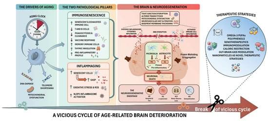
Mitochondria in Alzheimer’s Disease Pathogenesis

Alzheimer’s disease (AD) is a progressive and incurable neurodegenerative disorder that primarily affects persons aged 65 years and above. It causes dementia with memory loss and deterioration in thinking and language skills. AD is characterized by specific pathology resulting from the accumulation in the brain of extracellular plaques of amyloid-β and intracellular tangles of phosphorylated tau. The importance of mitochondrial dysfunction in AD pathogenesis, while previously underrecognized, is now more and more appreciated. Mitochondria are an essential organelle involved in cellular bioenergetics and signaling pathways. Mitochondrial processes crucial for synaptic activity such as mitophagy, mitochondrial trafficking, mitochondrial fission, and mitochondrial fusion are dysregulated in the AD brain. Excess fission and fragmentation yield mitochondria with low energy production.

Rheumatoid Factor: Diagnostic and Prognostic Performance and Therapeutic Implications in Rheumatoid Arthritis

Rheumatoid factor (RF) is the first autoantibody identified in rheumatoid arthritis (RA) which targets the fragment crystallizable (Fc) region of immunoglobulin (Ig) G. Although IgM isotype is predominant, other Ig isotypes, including IgG and IgA, also exist. While RF is not specific to RA, it remains a valuable serological test for diagnosing the disease, as evidenced by its inclusion in the 2010 classification criteria for RA based on elevated serum RF levels. RF is also associated with RA severity, including joint damage and extra-articular manifestations, serving as a poor prognostic factor and aiding in the identification of difficult-to-treat RA. Recent studies have demonstrated that high serum RF levels are associated with a reduced response to tumor necrosis factor (TNF) inhibitors.

How a chemical reaction triggers brain inflammation in Alzheimer’s disease

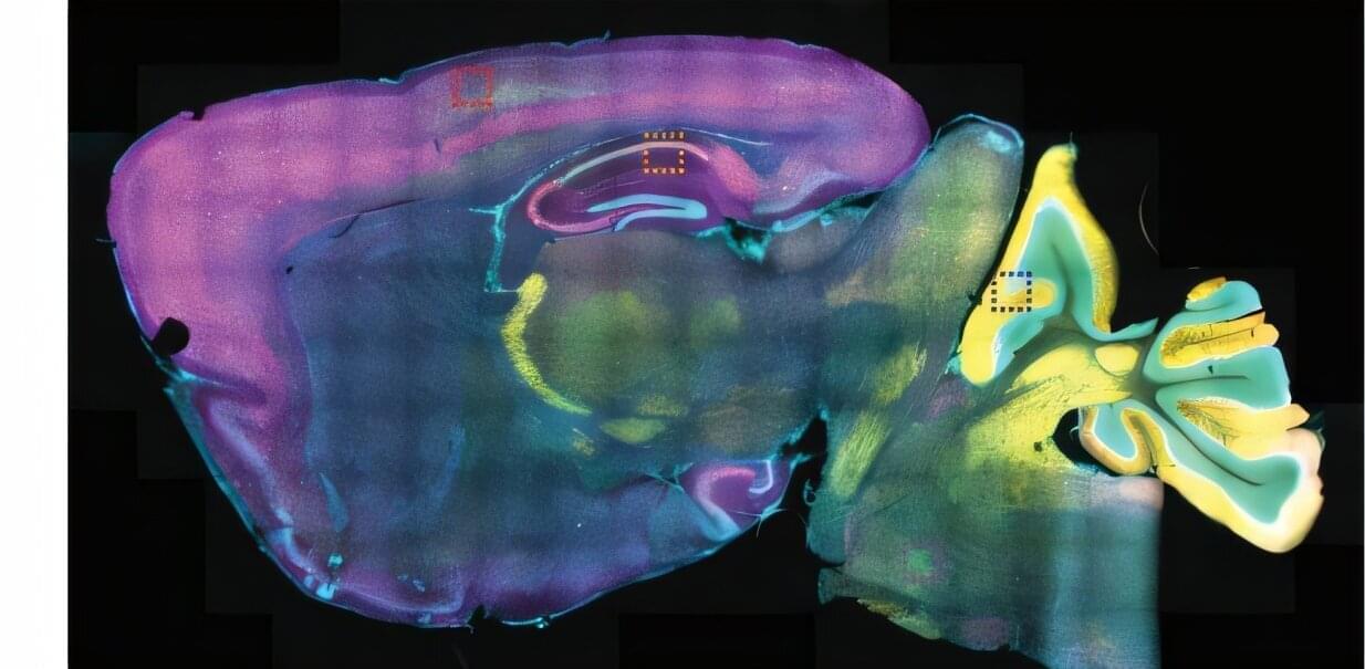
The brain has its own immune system, which detects threats and mounts a defense. A growing body of evidence has shown that in Alzheimer’s disease, those immune cells are chronically overactivated, causing inflammation that damages the connections between brain cells.

Now, in a preclinical study using human Alzheimer’s brain cells, scientists at Scripps Research have identified a molecular switch—and potential drug target—responsible for driving that chronic inflammation.

The research, published in Cell Chemical Biology on April 23, 2026, centers on a protein called STING, which normally functions as part of the immune system’s early-warning system. In the brains of people with Alzheimer’s, the team discovered that STING undergoes a chemical modification known as S-nitrosylation (or SNO, a reaction involving sulfur, oxygen and nitrogen) that promotes its overactivation. Blocking this chemical change to STING in a mouse model of the disease decreased neuroinflammation.

3D Recordings of Swimming Algae

Measurements of the 3D fluid flow around a swimming microorganism could help researchers better understand the swimming dynamics of such microbes.

Swimming microorganisms set up complex fluid flows that affect their ability to feed and communicate. Using advanced holographic methods, researchers have now imaged the entire 3D flow field around a swimming alga, revealing vortex rings that help propel the organism [1]. The researchers hope that the experiments will lead to improvements in measuring the energy expenditure and swimming strategies of a wide range of microorganisms.

The single-cell alga Chlamydomonas reinhardtii swims in a “breaststroke” style by beating its flagella—two hair-like appendages located at the front of its body—cycling 50 times per second. The flagella propel the organism forward while creating a surrounding fluid flow field that influences nutrient uptake and allows the organism to detect predators or mates. “The flow field generated by a swimming microorganism is one of its most fundamental characteristics,” says Xiang Cheng of the University of Minnesota. But he says that previous experiments have only captured partial details of this field, such as vortices to the left and right of the swimming organism. Researchers have speculated that these vortices might be connected in a larger coherent 3D flow pattern, but experiments have been unable to resolve such structures.

High-resolution imaging shines light on nanoscale nuclear organization

Researchers at the Indian Institute of Science (IISc) have implemented an advanced microscopy technique to visualize multiple biomolecules inside the nucleus of a cancer cell simultaneously at incredibly high resolution. The biomolecules they visualized include critical components of the cell’s transcription machinery and proteins that provide structural support to the nucleus—providing one of the first detailed maps of nuclear organization.

The human body is composed of trillions of cells. Each cell is an intricately organized meshwork of millions of proteins, nucleic acids, and many other molecules vital for the cell’s health. “Building novel technologies to visualize many biomolecules in individual cells is crucial to push the boundaries of biological research,” says Mahipal Ganji, Assistant Professor at the Department of Biochemistry (BC) and corresponding author of the study published in Nature Communications. Conventional imaging techniques, however, allow scientists to visualize only two or three biomolecules in each cell at a time.
In the study, the researchers turned to a microscopy technique called DNA-Points Accumulation for Imaging in Nanoscale Topography (DNA-PAINT), which allows for the visualization of biomolecules inside cells at incredible detail—far beyond the limits of conventional microscopes. It uses small fluorescent DNA fragments or tags that briefly attach to specific targets inside the cell and light up like tiny, blinking signals when a laser beam is shined on them.

Light near surface of ultra-thin optical fibers can sort twisted nanoparticles

Many important objects in the world can be divided into two categories based on their chirality or handedness, including molecules important for life such as amino acids. Such chiral objects (formally defined as objects which are not identical to their mirror images) are often characterized by a structure which twists in a given direction.

An everyday example of a chiral object is a screw. A right-handed screw moves into a material when rotated clockwise, but its mirror image (i.e., a left-handed screw) moves out.

Just as right-and left-handed screws behave differently when turned, chiral particles behave differently when exposed to light with a circular polarization. This fact allows them to be sorted in principle, which is expected to be important for applications such as drug development, where the handedness of a chiral molecule determines how it interacts with biological systems.

Re-engineered human cells boost gene-editing particle potency across multiple delivery systems

Gene editing has emerged as a powerful approach for targeting the genetic causes of disease, but getting the editing machinery into the right cells efficiently, safely, and at the scale needed for therapies remains one of the biggest set of challenges in the field.

Among the leading delivery vehicles are engineered virus-like particles, which resemble viruses—and share their knack for entering human cells—but carry no viral genes. Scientists load them with gene editing tools and use them to make precise changes in targeted cells.

Most efforts to improve these particles have focused on redesigning the particles themselves. A new study led by Valhalla Fellow at Whitehead Institute, Aditya Raguram and lab technician Diana Ly, focuses instead on the human cells that produce them.

/* */