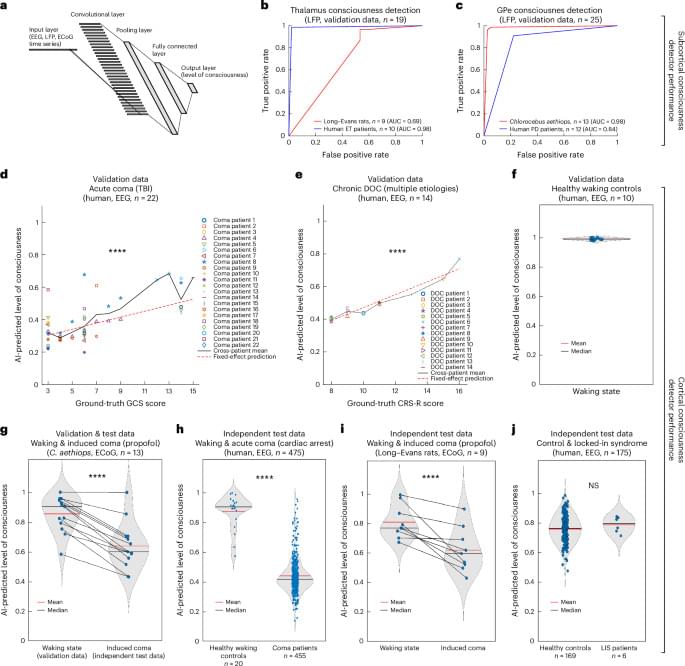
Toggle light / dark theme

A new study from Northwestern Medicine reports that, much like a conductor harmonizes various instruments in an orchestra to create a symphony, breathing synchronizes hippocampal brain waves to enhance memory during sleep.

This is the first time breathing rhythms during sleep have been linked to these hippocampal brain waves — called slow waves, spindles, and ripples — in humans. Scientists knew these waves were linked to memory but their underlying driver was unknown.

“To strengthen memories, three special neural oscillations emerge and synchronize in the hippocampus during sleep, but they were thought to come and go at random times,” said senior study author Christina Zelano, professor of neurology at Northwestern University.

A research team led by Prof. Nie Guangjun from the National Center for Nanoscience and Technology (NCNST) of the Chinese Academy of Sciences (CAS) and collaborators have demonstrated a tumor membrane antigens-based nanovaccine derived from liposomal doxorubicin treated tumor tissues, which is efficacious in inducing a potent immunological defense against tumors. The study is published online in Cell Reports Medicine.

For solid tumor surgeries, challenges remain in postoperative tumor recurrence and metastasis. The correlation between postoperative tumor recurrence and metastasis and the host’s antitumor immune status is well-established. Personalized cancer vaccines, using the patient’s own tumor as an antigen source, stimulate a robust immune response that is efficacious in eliminating residual neoplastic foci following as well as in targeting metastatic lesions at a distance, significantly reducing the risk of postoperative tumor recurrence and metastasis.

The efficacy of autologous tumor in has been limited by their weak immunogenicity. The tumor contains tumor-presented antigens and associated antigens, which can be developed into a personalized antigen library that more accurately reflects the expression of tumor antigens. Vaccines based on autologous tumor cell membrane antigens have been developed.

The all-in-one optical fiber spectrometer offers a compact microscale design with performance on par with traditional laboratory-based systems.

Miniaturized spectroscopy systems capable of detecting trace concentrations at parts-per-billion (ppb) levels are critical for applications such as environmental monitoring, industrial process control, and biomedical diagnostics.

However, conventional bench-top spectroscopy systems are often too large, complex, and impractical for use in confined spaces. Traditional laser spectroscopy techniques rely on bulky components—including light sources, mirrors, detectors, and gas cells—to measure light absorption or scattering. This makes them unsuitable for minimally invasive applications, such as intravascular diagnostics, where compactness and precision are essential.

Researchers at Australia’s Monash University are using a common medicine cabinet antiseptic in unique battery chemistry that could soon power drones and other electric aircraft, according to a school news release.

The team is tapping Betadine, a common brand name for a topical medication used to treat cuts and other wounds, in research garnering surprising results.

“… We found a way to accelerate the charge and discharge rates, making them a viable battery option for real-world heavy-duty use,” paper first author and doctoral student Maleesha Nishshanke said in the release.

What if robots could work together like ants to move objects, clear blockages, and guide living creatures? Discover more!

Scientists at Hanyang University in Seoul, South Korea, have developed small magnetic robots that work together in swarms to perform complex tasks, such as moving and lifting objects much more significant than themselves. These microrobot swarms, controlled by a rotating magnetic field, can be used in challenging environments, offering solutions for tasks like minimally invasive treatments for clogged arteries and guiding small organisms.

The researchers tested how microrobot swarms with different configurations performed various tasks. They discovered that swarms with a high aspect ratio could climb obstacles five times higher than a single robot’s body length and throw themselves over them. In another demonstration, a swarm of 1,000 microrobots formed a raft on water, surrounding a pill 2,000 times heavier than a single robot, allowing the swarm to transport the drug through the liquid. On land, a swarm moved cargo 350 times heavier than each robot, while another swarm unclogged tubes resembling blocked blood vessels. Using spinning and orbital dragging motions, the team also developed a system where robot swarms could guide the movements of small organisms.

Scientists have discovered a remarkable way to destroy cancer cells. A study published last year found stimulating aminocyanine molecules with near-infrared light caused them to vibrate in sync, enough to break apart the membranes of cancer cells.

Aminocyanine molecules are already used in bioimaging as synthetic dyes. Commonly used in low doses to detect cancer, they stay stable in water and are very good at attaching themselves to the outside of cells.

The research team from Rice University, Texas A&M University, and the University of Texas, said their approach is a marked improvement over another kind of cancer-killing molecular machine previously developed, called Feringa-type motors, which could also break the structures of problematic cells.

The evidence presented by Boldrini et al2 was considered resilient and convincing because the authors ensured that the samples were taken from healthy individuals using more biological parameters, such as angiogenesis and change in volume of DG, for a suitable comparison. They also employed unbiased stereology, which is the gold standard for counting number of neurons.2

The contrary results presented by the Sorrels study and Boldrini study highlight the existing ambiguity regarding the concept of neurogenesis in adult humans. Both studies employed reasonably similar immunohistological methods and included many of the same neurogenesis markers, yet contrasting results were observed. The study by Boldrini et al examined samples from humans aged 14 to 79 years, finding more than a thousand cells in each part of the DG, which was in stark contrast to what was observed by Sorrells et al, who found very few cells in the neurogenic niche of subjects in the same age range. Even if we consider that some discrepancies in numbers might arise due to the difference in counting methods (or subjective reasons), such marked and obvious disparity is perplexing. Sorrels et al justified their findings, noting the limitation that relying solely on the presence of markers might cause glial lineage cells to be identified as neuronal lineage cells. However, the authors stated that they used additional methods for confirmation, including transmission electron microscopy (TEM)-immunogold and in-situ hybridization. The relative paucity of any type of progenitor or immature cells, including glia in the neurogenic niche of DG in their study, remains unexplained.

Kempermann et al7 expressed skepticism regarding the negative findings of Sorrells et al, naming the postmortem interval, the lack of known status regarding neuropsychiatric disease or chronic ailment, and using patients with epilepsy as key factors of concern.7 Kempermann et al argued that, in severe epilepsy, destruction of the neurogenic niche is an explicit possibility, and that, in some cases, epilepsy could be the reason for the lack of neurogenesis.7 Additionally, Kempermann et al also criticized the use of 10% formalin in some of the samples due its potential to mask the expression of proteins.7 However, Sorrells et al clearly mentioned that they performed appropriate antigen retrieval for the selected sections.1 Kempermann et al7 suggested that dependence on the protein markers to denote neurogenesis could be an erroneous approach and that some of the markers, such as DCX, are known for fast degradation. Additionally, the presence of DCX-negative immature neurons and high inter-individual variation in expression of DCX in humans, which was also reflected in the data presented by Boldrini et al, are not unusual.7 The use of fluorescence markers also was presented as a caveat by Kempermann et al7 because it is prone to fade away and might give rise to false negative impression.

Scientists from the National University of Singapore (NUS) have developed a highly effective and general molecular design that enables an enhancement in radioluminescence within organometallic scintillators by more than three orders of magnitude. This enhancement harnesses X-ray-induced triplet exciton recycling within lanthanide metal complexes.

Detection of ionizing radiation is crucial in diverse fields, such as medical radiography, and astronomy. As a result, significant efforts have been dedicated to the development of luminescent materials that respond to X-rays.

However, current high-performance scintillators are almost exclusively limited to ceramic and perovskite materials, which face issues such as complex manufacturing processes, environmental toxicity, self-absorption and stability problems.

The company behind Oreo cookies has, by its own admission, been quietly creating new flavors using machine learning.

As the Wall Street Journal reports, Mondelez — the processed food behemoth that manufactures Oreos, Chips Ahoy, Clif Bars, and other popular snacks — has developed a new AI tool to dream up new flavors for its brands.

Used in more than 70 of the company’s products, the company says the machine learning tool is different from generative AI tools like ChatGPT and more akin to the drug discovery algorithms used by pharmaceutical companies to find and test new medications rapidly. Thus far the tool, created with the help of the software consultant Fourkind, has created products like the “Gluten Free Golden Oreo” and updated Chips Ahoy’s classic recipe, per the WSJ.