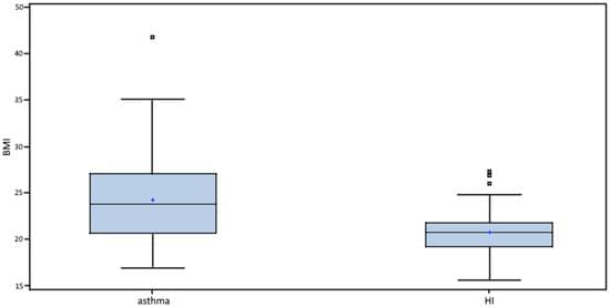
Our ability to use mathematical modelling is accelerating breakthrough discoveries in health care and biotechnology.
The cancer vaccine project is the latest indication of Amazon’s growing interest in the healthcare sector.
Distinct neuron types in the auditory organ are necessary for encoding different features of sound and relaying them to the brain. Researchers at Karolinska Institutet provide evidence of an early, neuronal activity-independent, emergence of the different subtypes of auditory neurons, prior to birth in mice. The findings have recently been published in Nature Communications.
Previous studies have provided ambiguous results on whether the different subtypes of auditory neurons emerge during prenatal or postnatal development, with in the latter case, a possible role of neuronal activity in generating their diversity. In this new study, researchers demonstrate that the fate of auditory neuron subtypes is under genetic control in the prenatal period, and reveal the complex molecular networks controlling their genesis.
The tiny crabs, which are about half a millimeter wide, can bend, twist, crawl, walk, turn, and even leap. Additionally, the scientists created millimeter-sized robots that resemble inchworms, crickets, and beetles. The study is experimental at this time, but the researchers think their technique might move the field closer to developing tiny robots that can carry out useful tasks in small, cramped areas.
The study was recently published in the journal Science Robotics. The same team also unveiled a winged microprocessor in September of last year; it was the tiniest flying object ever created by humans (published on the cover of Nature).
“Robotics is an exciting field of research, and the development of microscale robots is a fun topic for academic exploration,” said John A. Rogers, who led the experimental work. “You might imagine micro-robots as agents to repair or assemble small structures or machines in industry or as surgical assistants to clear clogged arteries, to stop internal bleeding or to eliminate cancerous tumors — all in minimally invasive procedures.”
Under a microscope, mammalian tissues reveal their intricate and elegant architectures. But if you look at the same tissue after tumour formation, you will see bedlam. Itai Yanai, a computational biologist at New York University’s Grossman School of Medicine in New York City, is trying to find order in this chaos. “There is a particular logic to how things are arranged, and spatial transcriptomics is helping us see that,” he says.
‘Spatial transcriptomics’ is a blanket term covering more than a dozen techniques for charting genome-scale gene-expression patterns in tissue samples, developed to complement single-cell RNA-sequencing techniques. Yet these single-cell sequencing methods have a downside — they can rapidly profile the messenger RNA content (or transcriptome) of large numbers of individual cells, but generally require physical disruption of the original tissue, which sacrifices crucial information about how cells are organized and can alter them in ways that might muddy later analyses. Immunologist Ido Amit at the Weizmann Institute of Science in Rehovot, Israel, says that such experiments would sometimes leave his group questioning their results. “Is this really the in situ state, or are we just looking at something which is either not a major [factor] or even not real at all?”
By contrast, spatial transcriptomics allows researchers to study gene expression in intact samples, opening frontiers in cancer research and revealing previously inaccessible biology of otherwise well-characterized tissues. The resulting ‘atlases’ of spatial information can tell scientists which cells make up each tissue, how they are organized and how they communicate. But compiling those atlases isn’t easy, because methods for spatial transcriptomics generally represent a tension between two competing goals: broader transcriptome coverage and tighter spatial resolution. Developments in experimental and computational methods are now helping researchers to balance those aims — and improving cellular resolution in the process.
Nematodes, a specific sort of microscopic worm, have been proven by Osaka University researchers to be capable of killing cancer cells, according to Interesting Engineering and SciTechDaily.
The study titled “Nematode surface functionalization with hydrogel sheaths tailored in situ” by Wildan Mubarok, Masaki Nakahata, Masaru Kojima and Shinji Sakai showed that Hydrogel-based “sheaths” that can be further modified to transport useful cargo (cancer-killing substances) could be applied to these worms as a coating.
View insights.
Often when Dr. Thomas Valley sees a new patient in the intensive care unit at Michigan Medicine in Ann Arbor, he clamps a pulse oximeter on their finger – one of the many devices he uses to gauge their health and what course of care they might require, whether they are a child having seizures, a teenage car accident victim or an older person with Covid-19.
But recently, Valley, an assistant professor in the University of Michigan’s Division of Pulmonary and Critical Care, realized first-hand that the small device may yield less accurate oxygen readings in patients with dark skin.
One end of the device sends light through the finger while a sensor on the other side receives this light and uses it to detect the color of your blood; bright red blood is highly oxygenated, while blue or purplish blood is less. If the device isn’t calibrated for darker skin tones, the pigmentation of the skin could affect how the light is absorbed by the sensor, leading to flawed oxygen readings.
Researchers from North Carolina State University have developed a new approach to federated learning that allows them to develop accurate artificial intelligence (AI) models more quickly and accurately. The work focuses on a longstanding problem in federated learning that occurs when there is significant heterogeneity in the various datasets being used to train the AI.
Federated learning is an AI training technique that allows AI systems to improve their performance by drawing on multiple sets of data without compromising the privacy of that data. For example, federated learning could be used to draw on privileged patient data from multiple hospitals in order to improve diagnostic AI tools, without the hospitals having access to data on each other’s patients.
Federated learning is a form of machine learning involving multiple devices, called clients. The clients and a centralized server all start with a basic model designed to solve a specific problem. From that starting point, each of the clients then trains its local model using its own data, modifying the model to improve its performance. The clients then send these “updates” to the centralized server. The centralized server draws on these updates to create a hybrid model, with the goal of having the hybrid model perform better than any of the clients on their own. The central server then sends this hybrid model back to each of the clients. This process is repeated until the system’s performance has been optimized or reaches an agreed-upon level of accuracy.
A new King’s-led study, published in the Proceedings of the National Academy of Sciences, has found that a single factor (a protein coding gene known as Sox8) can make non-ear cells adopt ear character during embryo development. The findings not only demonstrate how cell fate decisions are regulated in the embryo but may also inform reprogramming and regenerative strategies for the ear developmental malformations.
Responsible for the sense of hearing and balance, the inner ear is critically important for communication with the environment. In humans, developmental malformations of the ear have life-long consequences, while age-related hearing defects affect a large proportion of the population. Currently, there are no therapies that involve biological approaches—only hearing aids or cochlear implants, as how the ear normally develops is not fully understood and many of the controlling factors are poorly characterized.
Researchers from the Faculty of Dentistry, Oral and Craniofacial Sciences at King’s, in collaboration with colleagues from the Francis Crick Institute, explored the earliest steps in ear development to determine what causes cells to become ear cells, and what makes them different from cells which form other sense organs.