Toggle light / dark theme

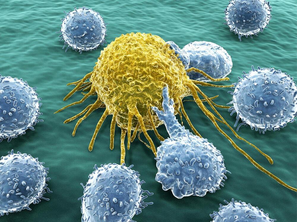
Nanoengineers at the University of California San Diego have developed immune cell-mimicking nanoparticles that target inflammation in the lungs and deliver drugs directly where they’re needed. As a proof of concept, the researchers filled the nanoparticles with the drug dexamethasone and administered them to mice with inflamed lung tissue. Inflammation was completely treated in mice given the nanoparticles, at a drug concentration where standard delivery methods did not have any efficacy.

The researchers reported their findings in Science Advances on June 16.

What’s special about these is that they are coated in a cell membrane that’s been genetically engineered to look for and bind to inflamed . They are the latest in the line of so-called cell membrane-coated nanoparticles that have been developed by the lab of UC San Diego nanoengineering professor Liangfang Zhang. His lab has previously used cell membrane-coated nanoparticles to absorb toxins produced by MRSA; treat sepsis; and train the immune system to fight cancer. But while these previous cell membranes were naturally derived from the body’s , the cell membranes used to coat this dexamethasone-filled nanoparticle were not.

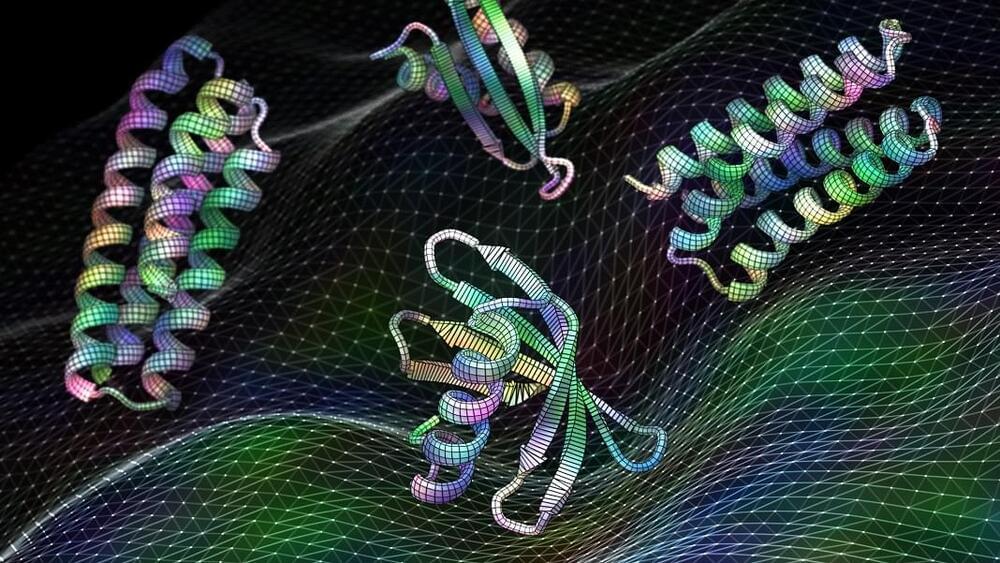
Check out my short video in which I explain some super exciting research in the area of nanotechnology: de novo protein lattices! I specifically discuss a journal article by Ben-Sasson et al. titled “Design of biologically active binary protein 2D materials”.


Here, I explain an exciting nanotechnology paper “Design of biologically active binary protein 2D materials” (https://doi.org/10.1038/s41586-020-03120-8).

Though I am not involved in this particular research myself, I have worked in adjacent areas such as de novo engineering of aggregating antimicrobial peptides, synthetic biology, nanotechnology-based tools for neuroscience, and gene therapy. I am endlessly fascinated by this kind of computationally driven de novo protein design and would love to incorporate it in my own research at some point in the future.

Circa 2015


Coatings that attract water (hydrophilic) are useful for anti-fogging applications6; any liquid water spreads out into a thin film thereby maintaining transparency. This is more favorable than using hydrophobic surfaces for anti-fogging as this requires a surface to be tilted for the droplets to roll off and transparency be maintained. Hydrophilic surfaces can also be used for self-cleaning7. Previous examples of superhydrophilic surfaces include the use of polymer–nanoparticle coatings8,9,10,11 however mechanical durability was not investigated.

Coatings with surface tensions lower than that of water (72 mN m–1) but higher than that of oils12 (20–30 mN m–1) will attract oils (oleophilic) but repel water and can be used to create oil–water separators13,14,15. When applied to a porous substrate, the coating will allow the passage of oil but block the passage of water, resulting in their separation. In addition, their water repellency also makes them ideal for self-cleaning4,16 and anti-icing17,18,19 applications. Anti-icing surfaces are typically superhydrophobic as supercooled droplets of water are able to roll off the cold surface before freezing and any ice formed is weakly adhered compared to hydrophilic surfaces due to an air cushion18,20.

Coatings with lower surface tensions (∼ 20 mN m–1 or less) will repel both oil (oleophobic) and water and are useful for anti-fouling such as in medical and transport applications, where both the oil-repellency and nanostructuring are of importance21,22,23,24,25,26,27. Previous work was not suitable for such applications as either the durability28 or oil-repellency29 was not optimal. The oil repellency also makes these surfaces ideal for anti-smudge applications30,31 where the oils from fingers are not deposited onto the surface and the surface remains clear. The water repellency means these coatings can also be used in self-cleaning and anti-icing applications.

Circa 2020


Researchers at UC Berkeley have developed a rapid test for SARS-CoV-2 that uses an enzyme to cleave viral RNA, initiating a fluorescent signal that can be detected using a smartphone camera, and which can provide a quantitative measurement of the level of viral particles in the sample. The test produce a result in as little as 30 minutes and does not require bulky or expensive laboratory equipment.

Rapid testing is key to measuring and stopping the spread of COVID-19, but current tests, such as PCR, are time consuming and require expensive laboratory equipment, creating a bottleneck in obtaining results. Researchers have been developing alternatives, and this latest technology was rapidly repurposed when the pandemic began. Originally intended to detect HIV in blood samples, the Berkeley researchers have pivoted to allow the device to detect SARS-CoV-2 in nasal swab samples.

Stimulation of the nervous system with neurotechnology has opened up new avenues for treating human disorders, such as prosthetic arms and legs that restore the sense of touch in amputees, prosthetic fingertips that provide detailed sensory feedback with varying touch resolution, and intraneural stimulation to help the blind by giving sensations of sight.

Scientists in a European collaboration have shown that optic nerve stimulation is a promising neurotechnology to help the blind, with the constraint that current technology has the capacity of providing only simple visual signals.

Nevertheless, the scientists’ vision (no pun intended) is to design these simple visual signals to be meaningful in assisting the blind with daily living. Optic nerve stimulation also avoids invasive procedures like directly stimulating the brain’s visual cortex. But how does one go about optimizing stimulation of the optic nerve to produce consistent and meaningful visual sensations?

Now, the results of a collaboration between EPFL, Scuola Superiore Sant’Anna and Scuola Internazionale Superiore di Studi Avanzati, published today in Patterns, show that a new stimulation protocol of the optic nerve is a promising way for developing personalized visual signals to help the blind–that also take into account signals from the visual cortex. The protocol has been tested for the moment on artificial neural networks known to simulate the entire visual system, called convolutional neural networks (CNN) usually used in computer vision for detecting and classifying objects. The scientists also performed psychophysical tests on ten healthy subjects that imitate what one would see from optic nerve stimulation, showing that successful object identification is compatible with results obtained from the CNN.

“We are not just trying to stimulate the optic nerve to elicit a visual perception,” explains Simone Romeni, EPFL scientist and first author of the study. “We are developing a way to optimize stimulation protocols that takes into account how the entire visual system responds to optic nerve stimulation.”

Hugh Herr is building the next generation of bionic limbs, robotic prosthetics inspired by nature’s own designs. Herr lost both legs in a climbing accident 30 years ago; now, as the head of the MIT Media Lab’s Biomechatronics group, he shows his incredible technology with the help of ballroom dancer Adrianne Haslet-Davis, who lost her left leg in the 2013 Boston Marathon bombing.

Automated data searches and new customized patient care are the future of cancer treatment.


Each day information floods into every cancer clinic. Oncologists are scrambling for new ways to tap it to deliver the best of modern cancer care.

This article was produced by Hackensack Meridian Health in partnership with Scientific American Custom Media, a division separate from the magazine’s board of editors.