Toggle light / dark theme

Phage therapy case reveals hidden antibodies can block treatment of drug-resistant infections

A new treatment for patients with life-threatening infectious diseases is being pioneered in Melbourne by researchers at The Alfred and Monash University. VICPhage, a clinical partnership between The Alfred and Monash, is one of the first in Australia to offer end-to-end capacity in phage therapy to treat some of the most challenging infections.

It involves injecting a patient with viruses called bacteriophages, or phages for short, to kill bacterial infections that have not responded to other treatments.

Professor Anton Peleg, Director of the Department of Infectious Diseases at The Alfred and Monash University and the Center to Impact AMR at Monash University, is co-lead of VICPhage and senior author of a new paper published in Nature Medicine.

How parasites exit host cells

After infecting host cells and reproducing, the parasite life cycle requires them to egress so that they can move to the next host. Past studies on the genes required for this process have been conducted but show conflicting results.

The methodology of past studies often involved opening the host cells during the screening process. Consequently, researchers were unable to reliably identify when mutations prevent parasites from egressing.

To avoid the same limitations, the team used an in vivo approach to screen for essential genes instead.

“Our in vivo screen, based on CRISPR, identified for the first time that the MIC11 gene is essential for host cell membrane permeabilization and parasite egress.” Explains the lead author.

Further tests demonstrated that deleting the MIC11 gene led the parasites to be unable to rupture the host cell membrane. By incapacitating parasites in this way, they could no longer exit the host cells, majorly disrupting the parasite life cycle.

“We also found evidence that MIC11 interacts with PLP1, providing further evidence of MIC11’s crucial role,” explains the senior author. “PLP1 is another parasite protein that was already known to be essential for egress.” ScienceMission sciencenewshighlights.


Frontiers: Year 2020 this gene therapy in mice shows promise for als gene therapy in humans

Gene therapy is an emerging and powerful therapeutic tool to deliver functional genetic material to cells in order to correct a defective gene. During the past decades, several studies have demonstrated the potential of AAV-based gene therapies for the treatment of neurodegenerative diseases. While some clinical studies have failed to demonstrate therapeutic efficacy, the use of AAV as a delivery tool has demonstrated to be safe. Here, we discuss the past, current and future perspectives of gene therapies for neurodegenerative diseases. We also discuss the current advances on the newly emerging RNAi-based gene therapies which has been widely studied in preclinical model and recently also made it to the clinic.

Gene therapy is an emerging therapeutic tool used to deliver functional genetic material to cells in order to correct a defective gene. By delivering a copy of a therapeutic gene to affected cells, the product encoded by that gene [i.e., its messenger RNA (mRNA) and/or proteins] will be continuously synthesized within the cell, utilizing the cell’s own transcriptional and translational machinery (Porada et al., 2013). The main advantage of this technology is that it offers a potentially life-long therapeutic effect without the need for repeated administration. Gene therapy can be used to correct defective genes by introducing a functional copy of the gene, by silencing a mutant allele using RNA interference (RNAi), by introducing a disease-modifying gene, or by using gene-editing technology (Grimm and Kay, 2007; Dow et al., 2015; Saraiva et al., 2016).

Gene therapy vectors can be either viral or non-viral. Different physical and chemical systems can be applied to deliver therapeutic genes to cells without the need of a viral vector. Non-viral vectors have no size limitation for the therapeutic gene, generally have a low immunogenicity risk, and can be produced at relatively low costs (Nayerossadat et al., 2012). However, due to the fact that high therapeutic doses are required when using non-viral technologies, and the resulting gene expression is generally transient, most gene therapies now rely on viral vectors. Numerous viral vector types have been tested in clinic, including vaccinia, measles, vesicular stomatitis virus (VSV), polio, reovirus, adenovirus, lentivirus, γ-retrovirus, herpes simplex virus (HSV) and adeno-associated virus (AAV) (Lundstrom, 2018).

Objectively Measured Daytime Napping and All-Cause Mortality in Older Adults

Among older adults, longer and more frequent daytime napping, especially in the morning, was associated with higher AllCauseMortality, supporting wearable sleep assessment for risk evaluation.


Question Are objectively measured daytime nap characteristics, including duration, frequency, variability, and timing, associated with all-cause mortality among community-dwelling older adults?

Findings In this prospective cohort study of 1,338 adults aged 56 years or older, longer and more frequent daytime napping, as well as morning napping, were associated with higher all-cause mortality. Variability in nap duration was not associated with mortality.

Meaning The findings suggest longer and more frequent, particularly morning, napping may be a behavioral marker of increased mortality risk in late life, underscoring the potential clinical value of incorporating wearable device–based nap assessments into routine health monitoring.

Protein’s second role in inflammation could reshape treatment for Crohn’s, arthritis and heart disease

A protein long understood to drive inflammation by producing nitric oxide has a second, previously unknown role—it physically binds to another key protein inside cells to directly modulate the immune response. The discovery, published in Nature Metabolism, could open new routes to treating conditions such as cardiovascular disease, arthritis, Crohn’s and other inflammatory diseases.

When the immune system detects infection or injury, it triggers inflammation to fight back. That response is essential, but it must be carefully controlled. If it runs too hard for too long, it causes the tissue damage that underlies many chronic diseases. Understanding the molecular switches that regulate inflammation—and finding new ways to target them—is one of the biggest challenges in modern medicine.

Researchers from the University of Surrey and the University of Oxford have identified one such switch. They have shown that inducible nitric oxide synthase (iNOS)—a protein that produces nitric oxide during inflammation—can also bind directly to a second protein, IRG1, inside mitochondria. That physical interaction blocks IRG1 from producing itaconate, a metabolite that acts as a brake on the inflammatory response.

Abstract: Expanding clones, expanding aneurysms through macrophage-to-osteoclast differentiation:

Jessica A. Regan & Svati H. Shah Comment on Yonekawa et al.: https://doi.org/10.1172/JCI198708 aneurysm.


Address correspondence to: Jessica A. Regan, Duke Molecular Physiology Institute Duke University School of Medicine, 300 N. Duke Street, Carmichael Building, Durham, North Carolina, 27,701, USA. Email: [email protected].

Real-time impedance-based cell migration measurements with integrated electrodes on porous membranes for next generation microphysiological systems

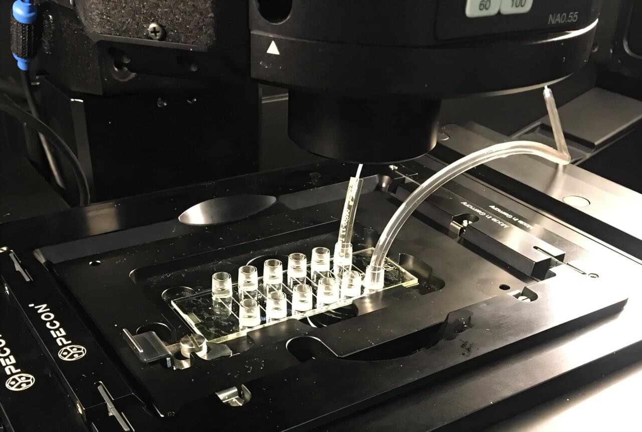
A new laboratory technique for measuring how quickly cells penetrate and pass through a porous membrane and reach the opposite side could help identify cancer cells with the greatest potential to spread in the human body.

The method relies on tiny electrodes placed on either side of an artificial membrane. The electrodes measure changes in electrical resistance as cells pass through the material. The most aggressive cancer cells pass through the membrane more rapidly than other cells.

The illustration depicts cells (green and blue) moving through a membrane (grey) studded with microelectrodes (gold rings).

🔗


We present a novel microfluidic device capable of electrically interrogating both surfaces of a porous membrane quantitatively and in real time using electrical impedance spectroscopy to monitor cell migration. This device holds patterned gold electrodes on both sides of the membrane, which enable independent impedance measurements on each side of the membrane. We introduce the term cross-over cell migration (CoCM) to describe this dual-sided approach, which allows precise monitoring of cells at their seeding location and as they move through a porous membrane. To ensure reliable tracking, we developed a normalization method, the CoCM index, that allows us to compare both membrane surfaces directly in real-time. Human renal carcinoma cells (786-O) were passively seeded in the device’s top microfluidic chamber, and we collected impedance data from both sides of the membrane surfaces simultaneously over a three-day period. These measurements successfully captured the onset and progression of cell migration across the membrane interface. We tracked the cells with fluorescence imaging in parallel to validate our impedance data. As cells appeared in focus on the bottom-side electrode surface, their numbers kept increasing over the course of our experiment. The CoCM index decreased by about 20% in the top chamber and increased by approximately 15% in the bottom chamber. Symmetrical CoCM index trends appeared after 40 h, consistent with the fluorescent images captured. Finally, we performed live-cell fluorescence assays to confirm post-experiment cell viability and to quantify migrated cells, further validating our CoCM platform measurements. This platform is a valuable tool not only for real-time and quantitative cell migration studies of cancer and other cells in bulk but also for future studies of single-cell migration processes.

This artificial retina doesn’t just aim to restore sight—it opens a hidden channel of vision

The retina, the thin layer of tissue at the back of the eye, is made up of photoreceptor cells that convert visible light into electrical signals, which is essential for human vision. Some diseases, such as retinal degeneration, cause these photoreceptor cells to stop working, which results in blindness. Researchers at Yonsei University, the Institute for Basic Science (IBS) and other institutes in the Republic of Korea have recently developed a new artificial retina that could partly restore vision in people with damaged retinas.

The new device, introduced in a paper published in Nature Electronics, works by detecting near-infrared light and converting it into electrical signals, which stimulate another type of cells in the retina that are undamaged.

“Many people suffer from blindness due to retinal diseases that cause photoreceptor degeneration,” wrote Won Gi Chung, Inhea Jeong and their colleagues in their paper. “Electrical stimulation of retinal neurons can recreate the action potentials associated with seeing that are generated by these cells. We report a thin artificial retina that can be adhered to the epiretinal surface and can convert near-infrared (NIR) light into electrical stimuli that selectively stimulate ganglion cells.”

Subversion of kynurenine-induced AHR activation in CD8 T cells by kynureninase-expressing antigen-presenting cells

Giacomantonio et al. demonstrate an immunomodulatory role for a metabolic enzyme kynureninase in antigen-presenting cells (APCs). Kynureninase-expressing APCs degrade kynurenine, including that generated by cancer cells, and relieve kynurenine-induced immunosuppression in CD8+ T cells.

/* */