Toggle light / dark theme

The U.S. Department of Health and Human Services (HHS) has proposed updates to the Health Insurance Portability and Accountability Act of 1996 (HIPAA) to secure patients’ health data following a surge in massive healthcare data leaks.

These stricter cybersecurity rules, proposed by the HHS’ Office for Civil Rights (OCR) and expected to be published as a final rule within 60 days, would require healthcare organizations to encrypt protected health information (PHI), implement multifactor authentication, and segment their networks to make it harder for attackers to move laterally through them.

“In recent years, there has been an alarming growth in the number of breaches affecting 500 or more individuals reported to the Department, the overall number of individuals affected by such breaches, and the rampant escalation of cyberattacks using hacking and ransomware,” the HHS’ proposal says.

Research led by Zhejiang Agriculture and Forestry University in China has performed a metadata investigation into the presence of microplastics in humans. They report a concerning relationship between micro and nanoplastic (MNP) concentrations in damaged tissues and links with multiple health conditions.

Plastic usage soared from 1.5 million metric tons in the 1950s to nearly 390.7 million in 2021. With the increased use in came elevated microscopic plastic pollution circulating in soil and waterways, eventually accumulating in the environment, food webs and human tissues.

Consistent methods to pinpoint and quantify MNPs in human tissues are lacking. Reliable data linking MNPs to human diseases are necessary for assessing potential risks and developing mitigation measures.

Have you ever wondered how fast our brains work? Well, scientists have recently quantified the brain’s speed limit. They revealed that from sensory organs, the brain processes signals at only about 10 bits per second.

This speed is millions of times slower than the input rate, as the human body’s sensory systems gather data about the surrounding environment at a rate of a billion bits per second.

Earlier, Director of the Gamaleya National Research Center for Epidemiology and Microbiology Alexander Gintsburg told TASS that the vaccine’s pre-clinical trials had shown that it suppresses tumor development and potential metastases.


MOSCOW, December 15. /TASS/. Russia has developed its own mRNA vaccine against cancer, it will be distributed to patients free of charge, General Director of the Radiology Medical Research Center of the Russian Ministry of Health Andrey Kaprin has told Radio Rossiya.

The vaccine was developed in collaboration with several research centers. It is planned to launch it in general circulation in early 2025.

© Mikhail Sinitsyn/TASS

Love this interview with Erika Alden DeBenedictis on her work towards terraforming Mars with engineered microorganisms, her thoughts about how to develop new funding structures for biotechnology, and her ideas on finding a balance between standardizing practices across biotechnology and retaining customizability. #biotech #mars #future #research


A conversation with Astera resident Erika Alden DeBenedictis.

The secret to cellular youth may lie in maintaining a small nucleolus—a dense structure within the cell nucleus—according to investigators at Weill Cornell Medicine. These findings were uncovered in yeast, a model organism renowned for its role in making bread and beer, yet surprisingly similar to humans at the cellular level.

The study, published Nov. 25 in Nature Aging, may lead to new longevity treatments that could extend human lifespan. It also establishes a mortality timer that reveals how long a cell has left before it dies.

As people get older, they are more likely to develop health conditions, such as cancer, cardiovascular disease and neurodegenerative diseases.

Metabolic imaging is a noninvasive method that enables clinicians and scientists to study living cells using laser light, which can help them assess disease progression and treatment responses.

But light scatters when it shines into biological tissue, limiting how deep it can penetrate and hampering the resolution of captured images.

Now, MIT researchers have developed a new technique that more than doubles the usual depth limit of metabolic imaging. Their method also boosts imaging speeds, yielding richer and more detailed images.

This new technique does not require tissue to be preprocessed, such as by cutting it or staining it with dyes. Instead, a specialized laser illuminates deep into the tissue, causing certain intrinsic molecules within the cells and tissues to emit light. This eliminates the need to alter the tissue, providing a more natural and accurate representation of its structure and function.


MIT researchers developed a non-invasive imaging technique that enables laser light to penetrate deeper into living tissue, capturing sharper images of cells. This could help clinical biologists study disease progression and develop new medicines.

Decades of research have established that chronic stress—from money worries, job problems, family tensions, or other sources—causes chemical changes in the body. In a new study, researchers have identified biological changes induced by stress that may help explain how it could cause a tumor to spread, or metastasize.


To conduct the study, the researchers used two established methods for modeling stress in mice. One is designed to mimic exposure to constant, low-level, predictable stress. The other simulates intermittent, unpredictable, mild stress.

They used these methods to induce chronic stress in two different mouse models of breast cancer. In both models, when the mice were exposed to stress using either method, they had both larger mammary tumors and more lung metastases than mice not exposed to stress.

But a series of follow-up experiments strongly suggested that this increased tumor growth and metastasis wasn’t being driven by the effects of stress on cancer cells themselves.

Surprisingly, the organoids were still healthy when they returned from orbit a month later, but the cells had matured faster compared to identical organoids grown on Earth—they were closer to becoming adult neurons and were beginning to show signs of specialization. The results, which could shed light on potential neurological effects of space travel, were published on October 23, 2024, in Stem Cells Translational Medicine.

“The fact that these cells survived in space was a big surprise,” says co-senior author Jeanne Loring, PhD, professor emeritus in the Department of Molecular Medicine and founding director of the Center for Regenerative Medicine at Scripps Research. “This lays the groundwork for future experiments in space, in which we can include other parts of the brain that are affected by neurodegenerative disease.”

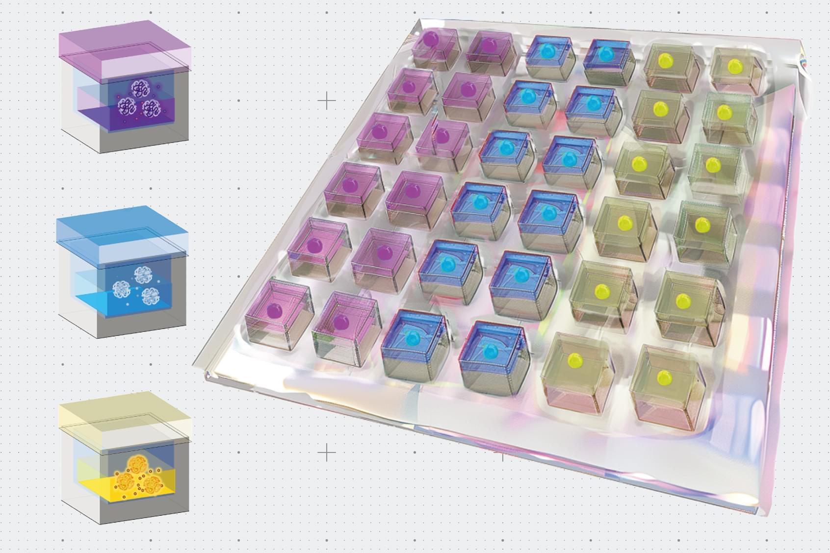
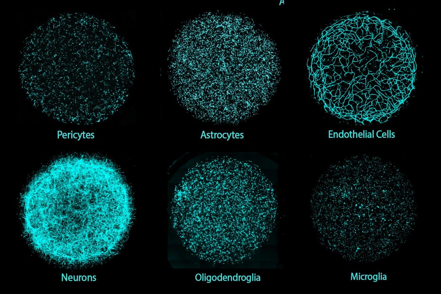
On Earth, the team used stem cells to create organoids consisting of either cortical or dopaminergic neurons, which are the neuronal populations impacted in multiple sclerosis and Parkinson’s disease—diseases that Loring has studied for decades. Some organoids also included microglia, a type of immune cell that is resident within the brain, to examine the impact of microgravity on inflammation.


Abstract. Research conducted on the International Space Station (ISS) in low-Earth orbit (LEO) has shown the effects of microgravity on multiple organs. To investigate the effects of microgravity on the central nervous system, we developed a unique organoid strategy for modeling specific regions of the brain that are affected by neurodegenerative diseases. We generated 3-dimensional human neural organoids from induced pluripotent stem cells (iPSCs) derived from individuals affected by primary progressive multiple sclerosis (PPMS) or Parkinson’s disease (PD) and non-symptomatic controls, by differentiating them toward cortical and dopaminergic fates, respectively, and combined them with isogenic microglia. The organoids were cultured for a month using a novel sealed cryovial culture method on the International Space Station (ISS) and a parallel set that remained on Earth. Live samples were returned to Earth for analysis by RNA expression and histology and were attached to culture dishes to enable neurite outgrowth. Our results show that both cortical and dopaminergic organoids cultured in LEO had lower levels of genes associated with cell proliferation and higher levels of maturation-associated genes, suggesting that the cells matured more quickly in LEO. This study is continuing with several more missions in order to understand the mechanisms underlying accelerated maturation and to investigate other neurological diseases. Our goal is to make use of the opportunity to study neural cells in LEO to better understand and treat neurodegenerative disease on Earth and to help ameliorate potentially adverse neurological effects of space travel.

Mount Sinai researchers discovered that harmine, a beta cell regenerative drug, may transform alpha cells into beta cells, offering scalable diabetes treatment options for millions.

Researchers and bioinformaticians at the Icahn School of Medicine at Mount Sinai have unveiled new insights into the mechanisms behind human beta cell regenerative drugs, offering a potential breakthrough for the over 500 million people worldwide living with diabetes. These findings, recently published in Cell Reports Medicine, could mark a significant step forward in diabetes treatment.

Diabetes occurs when pancreatic beta cells lose their ability to produce insulin, a hormone critical for maintaining healthy blood sugar levels. Despite significant advancements, there are still no widely scalable therapeutic solutions capable of addressing the global diabetes crisis.